

administration is urgently needed for the therapeutic application of EVs [1–3].
Thus, several noninvasive imaging methods have been developed for tracking EVs.
They basically employed labeling exogenous tracers which generate signals ranged
from visible light to radiation as EVs could be loaded with various bioactive
materials including tracers as well as drugs, nanoparticles, genetic materials such as
microRNAs [4, 5].
The loading methods of tracers can be roughly classified into two categories:
(1) A strategy that parent cells load tracers so that the later-secreted EVs carry the
targeted materials and (2) another strategy of direct labeling and decorating EVs.
Using the first strategy, several hybrid EVs loading magnetic, fluorescent and
therapeutic nanoparticles can be produced [6–8]. In addition, genetic modification
of the parent cells allows to produce sustainably the EVs that inherit fluorescence
proteins [9, 10]. In contrast, the second strategy is more flexible and simple for the
labeling as the direct labeling is feasible regardless of types of EVs obtained from
various cell types and conditions. Because several lipophilic drugs may interact
directly with lipid bilayers of EVs, the labeling can be performed under physiologic
condition by incubating lipophilic tracers and EVs.
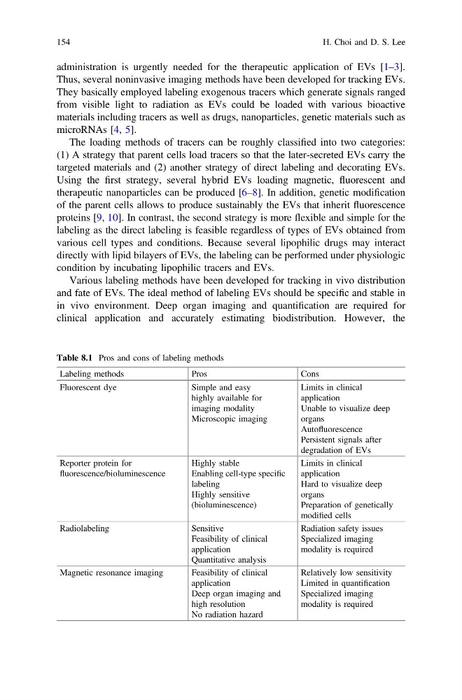
Various labeling methods have been developed for tracking in vivo distribution
and fate of EVs. The ideal method of labeling EVs should be specific and stable in
in vivo environment. Deep organ imaging and quantification are required for
clinical application and accurately estimating biodistribution. However, the
Table 8.1 Pros and cons of labeling methods
Labeling methods
Pros
Cons
Fluorescent dye
Simple and easy
highly available for
imaging modality
Microscopic imaging
Limits in clinical
application
Unable to visualize deep
organs
Autofluorescence
Persistent signals after
degradation of EVs
Reporter protein for
fluorescence/bioluminescence
Highly stable
Enabling cell-type specific
labeling
Highly sensitive
(bioluminescence)
Limits in clinical
application
Hard to visualize deep
organs
Preparation of genetically
modified cells
Radiolabeling
Sensitive
Feasibility of clinical
application
Quantitative analysis
Radiation safety issues
Specialized imaging
modality is required
Magnetic resonance imaging
Feasibility of clinical
application
Deep organ imaging and
high resolution
No radiation hazard
Relatively low sensitivity
Limited in quantification
Specialized imaging
modality is required
154
H. Choi and D. S. Lee
Thus, several noninvasive imaging methods have been developed for tracking EVs.
They basically employed labeling exogenous tracers which generate signals ranged
from visible light to radiation as EVs could be loaded with various bioactive
materials including tracers as well as drugs, nanoparticles, genetic materials such as
microRNAs [4, 5].
The loading methods of tracers can be roughly classified into two categories:
(1) A strategy that parent cells load tracers so that the later-secreted EVs carry the
targeted materials and (2) another strategy of direct labeling and decorating EVs.
Using the first strategy, several hybrid EVs loading magnetic, fluorescent and
therapeutic nanoparticles can be produced [6–8]. In addition, genetic modification
of the parent cells allows to produce sustainably the EVs that inherit fluorescence
proteins [9, 10]. In contrast, the second strategy is more flexible and simple for the
labeling as the direct labeling is feasible regardless of types of EVs obtained from
various cell types and conditions. Because several lipophilic drugs may interact
directly with lipid bilayers of EVs, the labeling can be performed under physiologic
condition by incubating lipophilic tracers and EVs.
Various labeling methods have been developed for tracking in vivo distribution
and fate of EVs. The ideal method of labeling EVs should be specific and stable in
in vivo environment. Deep organ imaging and quantification are required for
clinical application and accurately estimating biodistribution. However, the
Table 8.1 Pros and cons of labeling methods
Labeling methods
Pros
Cons
Fluorescent dye
Simple and easy
highly available for
imaging modality
Microscopic imaging
Limits in clinical
application
Unable to visualize deep
organs
Autofluorescence
Persistent signals after
degradation of EVs
Reporter protein for
fluorescence/bioluminescence
Highly stable
Enabling cell-type specific
labeling
Highly sensitive
(bioluminescence)
Limits in clinical
application
Hard to visualize deep
organs
Preparation of genetically
modified cells
Radiolabeling
Sensitive
Feasibility of clinical
application
Quantitative analysis
Radiation safety issues
Specialized imaging
modality is required
Magnetic resonance imaging
Feasibility of clinical
application
Deep organ imaging and
high resolution
No radiation hazard
Relatively low sensitivity
Limited in quantification
Specialized imaging
modality is required
154
H. Choi and D. S. Lee