

The Effect of Enzyme Addition on the Anaerobic Digestion …
125
3.5
3.7
3.9
4.1
4.3
4.5
4.7
4.9
5.1
5.3
5.5
0
1 0
2 0
3 0
4 0
5 0
pH
Time, days
Food Waste
Food Waste and
Lipase
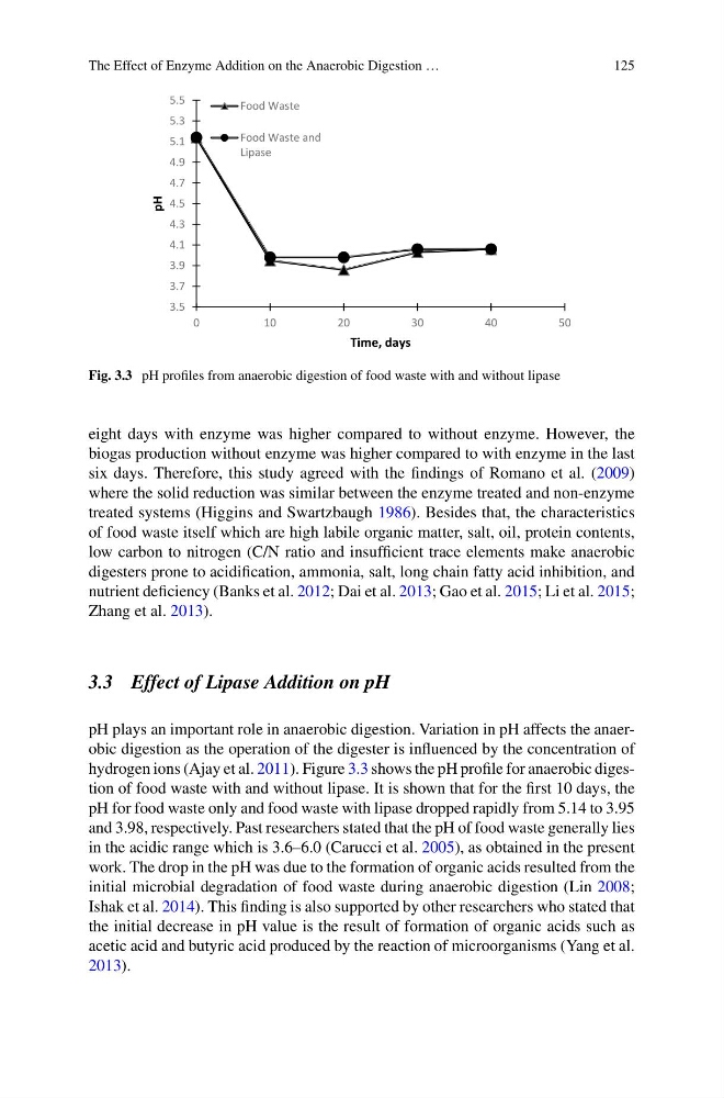
Fig. 3.3 pH profiles from anaerobic digestion of food waste with and without lipase
eight days with enzyme was higher compared to without enzyme. However, the
biogas production without enzyme was higher compared to with enzyme in the last
six days. Therefore, this study agreed with the findings of Romano et al. (2009)
where the solid reduction was similar between the enzyme treated and non-enzyme
treated systems (Higgins and Swartzbaugh 1986). Besides that, the characteristics
of food waste itself which are high labile organic matter, salt, oil, protein contents,
low carbon to nitrogen (C/N ratio and insufficient trace elements make anaerobic
digesters prone to acidification, ammonia, salt, long chain fatty acid inhibition, and
nutrient deficiency (Banks et al. 2012; Dai et al. 2013; Gao et al. 2015; Li et al. 2015;
Zhang et al. 2013).
3.3 Effect of Lipase Addition on pH
pH plays an important role in anaerobic digestion. Variation in pH affects the anaerobic digestion as the operation of the digester is influenced by the concentration of
hydrogen ions (Ajay et al. 2011). Figure 3.3 shows the pH profile for anaerobic digestion of food waste with and without lipase. It is shown that for the first 10 days, the
pH for food waste only and food waste with lipase dropped rapidly from 5.14 to 3.95
and 3.98, respectively. Past researchers stated that the pH of food waste generally lies
in the acidic range which is 3.6–6.0 (Carucci et al. 2005), as obtained in the present
work. The drop in the pH was due to the formation of organic acids resulted from the
initial microbial degradation of food waste during anaerobic digestion (Lin 2008;
Ishak et al. 2014). This finding is also supported by other researchers who stated that
the initial decrease in pH value is the result of formation of organic acids such as
acetic acid and butyric acid produced by the reaction of microorganisms (Yang et al.
2013).
125
3.5
3.7
3.9
4.1
4.3
4.5
4.7
4.9
5.1
5.3
5.5
0
1 0
2 0
3 0
4 0
5 0
pH
Time, days
Food Waste
Food Waste and
Lipase
Fig. 3.3 pH profiles from anaerobic digestion of food waste with and without lipase
eight days with enzyme was higher compared to without enzyme. However, the
biogas production without enzyme was higher compared to with enzyme in the last
six days. Therefore, this study agreed with the findings of Romano et al. (2009)
where the solid reduction was similar between the enzyme treated and non-enzyme
treated systems (Higgins and Swartzbaugh 1986). Besides that, the characteristics
of food waste itself which are high labile organic matter, salt, oil, protein contents,
low carbon to nitrogen (C/N ratio and insufficient trace elements make anaerobic
digesters prone to acidification, ammonia, salt, long chain fatty acid inhibition, and
nutrient deficiency (Banks et al. 2012; Dai et al. 2013; Gao et al. 2015; Li et al. 2015;
Zhang et al. 2013).
3.3 Effect of Lipase Addition on pH
pH plays an important role in anaerobic digestion. Variation in pH affects the anaerobic digestion as the operation of the digester is influenced by the concentration of
hydrogen ions (Ajay et al. 2011). Figure 3.3 shows the pH profile for anaerobic digestion of food waste with and without lipase. It is shown that for the first 10 days, the
pH for food waste only and food waste with lipase dropped rapidly from 5.14 to 3.95
and 3.98, respectively. Past researchers stated that the pH of food waste generally lies
in the acidic range which is 3.6–6.0 (Carucci et al. 2005), as obtained in the present
work. The drop in the pH was due to the formation of organic acids resulted from the
initial microbial degradation of food waste during anaerobic digestion (Lin 2008;
Ishak et al. 2014). This finding is also supported by other researchers who stated that
the initial decrease in pH value is the result of formation of organic acids such as
acetic acid and butyric acid produced by the reaction of microorganisms (Yang et al.
2013).