

7. Optional: Fragmentation of larger crystals, i.e., bigger than
about 2 μm for the smallest side. First add approximately
10 μL of glass beads (Research Products International) to a
1.5 mL Eppendorf tube and wash them with 500 μL of 20%
(v/v) ethanol, followed by washing with 500 μL dH 2 O. Then
the beads are equilibrated with about 30 μL of stabilizing
buffer (e.g., reservoir buffer). Pipette the collected crystals
into the Eppendorf tube with the glass beads. The crystals are
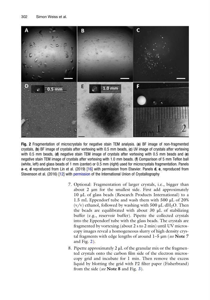
fragmented by vortexing (about 2 s to 2 min) until UV microscopy images reveal a homogeneous slurry of high density crystal fragments with edge lengths of around 1–5 μm (see Note 7
and Fig. 2).
8. Pipette approximately 2 μL of the granular mix or the fragmented crystals onto the carbon film side of the electron microscopy grid and incubate for 1 min. Then remove the excess
liquid by blotting the grid with P2 filter paper (Fisherbrand)
from the side (see Note 8 and Fig. 3).
Fig. 2 Fragmentation of microcrystals for negative stain TEM analysis. (a) BF image of non-fragmented
crystals, (b) BF image of crystals after vortexing with 0.5 mm beads, (c) UV image of crystals after vortexing
with 0.5 mm beads, (d) negative stain TEM image of crystals after vortexing with 0.5 mm beads and (e)
negative stain TEM image of crystals after vortexing with 1.0 mm beads. (f) Comparison of 5 mm Teflon ball
(white, left) and glass beads of 1 mm (center) or 0.5 mm (right) used for microcrystals fragmentation. Panels
a–c, d reproduced from Lin et al. (2019) [16] with permission from Elsevier. Panels d, e, reproduced from
Stevenson et al. (2016) [12] with permission of the International Union of Crystallography
302
Simon Weiss et al.
about 2 μm for the smallest side. First add approximately
10 μL of glass beads (Research Products International) to a
1.5 mL Eppendorf tube and wash them with 500 μL of 20%
(v/v) ethanol, followed by washing with 500 μL dH 2 O. Then
the beads are equilibrated with about 30 μL of stabilizing
buffer (e.g., reservoir buffer). Pipette the collected crystals
into the Eppendorf tube with the glass beads. The crystals are
fragmented by vortexing (about 2 s to 2 min) until UV microscopy images reveal a homogeneous slurry of high density crystal fragments with edge lengths of around 1–5 μm (see Note 7
and Fig. 2).
8. Pipette approximately 2 μL of the granular mix or the fragmented crystals onto the carbon film side of the electron microscopy grid and incubate for 1 min. Then remove the excess
liquid by blotting the grid with P2 filter paper (Fisherbrand)
from the side (see Note 8 and Fig. 3).
Fig. 2 Fragmentation of microcrystals for negative stain TEM analysis. (a) BF image of non-fragmented
crystals, (b) BF image of crystals after vortexing with 0.5 mm beads, (c) UV image of crystals after vortexing
with 0.5 mm beads, (d) negative stain TEM image of crystals after vortexing with 0.5 mm beads and (e)
negative stain TEM image of crystals after vortexing with 1.0 mm beads. (f) Comparison of 5 mm Teflon ball
(white, left) and glass beads of 1 mm (center) or 0.5 mm (right) used for microcrystals fragmentation. Panels
a–c, d reproduced from Lin et al. (2019) [16] with permission from Elsevier. Panels d, e, reproduced from
Stevenson et al. (2016) [12] with permission of the International Union of Crystallography
302
Simon Weiss et al.