

6. Optional: Vortex Mixer.
7. Optional: glass beads 0.5 mm or 1 mm.
3 Methods
1. Set up hanging drop vapor diffusion crystallization screens of
your target protein (see Note 1).
2. Identify conditions with granular aggregate and micro
or nanocrystals by screening the crystallization trays under a
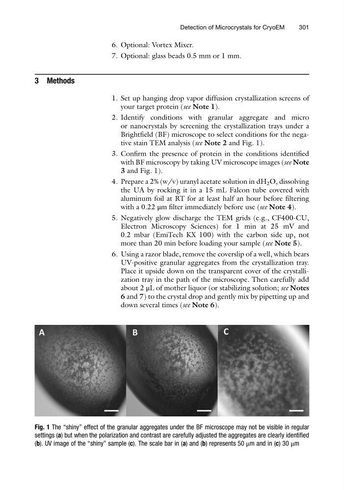
Brightfield (BF) microscope to select conditions for the negative stain TEM analysis (see Note 2 and Fig. 1).
3. Confirm the presence of protein in the conditions identified
with BF microscopy by taking UV microscope images (see Note
3 and Fig. 1).
4. Prepare a 2% (w/v) uranyl acetate solution in dH 2 O, dissolving
the UA by rocking it in a 15 mL Falcon tube covered with
aluminum foil at RT for at least half an hour before filtering
with a 0.22 μm filter immediately before use (see Note 4).
5. Negatively glow discharge the TEM grids (e.g., CF400-CU,
Electron Microscopy Sciences) for 1 min at 25 mV and
0.2 mbar (EmiTech KX 100) with the carbon side up, not
more than 20 min before loading your sample (see Note 5).
6. Using a razor blade, remove the coverslip of a well, which bears
UV-positive granular aggregates from the crystallization tray.
Place it upside down on the transparent cover of the crystallization tray in the path of the microscope. Then carefully add
about 2 μL of mother liquor (or stabilizing solution; see Notes
6 and 7) to the crystal drop and gently mix by pipetting up and
down several times (see Note 6).
Fig. 1 The “shiny” effect of the granular aggregates under the BF microscope may not be visible in regular
settings (a) but when the polarization and contrast are carefully adjusted the aggregates are clearly identified
(b). UV image of the “shiny” sample (c). The scale bar in (a) and (b) represents 50 μm and in (c) 30 μm
Detection of Microcrystals for CryoEM
301
7. Optional: glass beads 0.5 mm or 1 mm.
3 Methods
1. Set up hanging drop vapor diffusion crystallization screens of
your target protein (see Note 1).
2. Identify conditions with granular aggregate and micro
or nanocrystals by screening the crystallization trays under a
Brightfield (BF) microscope to select conditions for the negative stain TEM analysis (see Note 2 and Fig. 1).
3. Confirm the presence of protein in the conditions identified
with BF microscopy by taking UV microscope images (see Note
3 and Fig. 1).
4. Prepare a 2% (w/v) uranyl acetate solution in dH 2 O, dissolving
the UA by rocking it in a 15 mL Falcon tube covered with
aluminum foil at RT for at least half an hour before filtering
with a 0.22 μm filter immediately before use (see Note 4).
5. Negatively glow discharge the TEM grids (e.g., CF400-CU,
Electron Microscopy Sciences) for 1 min at 25 mV and
0.2 mbar (EmiTech KX 100) with the carbon side up, not
more than 20 min before loading your sample (see Note 5).
6. Using a razor blade, remove the coverslip of a well, which bears
UV-positive granular aggregates from the crystallization tray.
Place it upside down on the transparent cover of the crystallization tray in the path of the microscope. Then carefully add
about 2 μL of mother liquor (or stabilizing solution; see Notes
6 and 7) to the crystal drop and gently mix by pipetting up and
down several times (see Note 6).
Fig. 1 The “shiny” effect of the granular aggregates under the BF microscope may not be visible in regular
settings (a) but when the polarization and contrast are carefully adjusted the aggregates are clearly identified
(b). UV image of the “shiny” sample (c). The scale bar in (a) and (b) represents 50 μm and in (c) 30 μm
Detection of Microcrystals for CryoEM
301