

least one neighbor protein on each side, implying an overlap
between the boxes of adjacent particles, as shown by the blue
boxes in Fig. 1c. This also favors the smoothness of the alignment
parameters across the 2D crystal because of the natural correlations
existing among adjacent particles (proteins located close to each
other in the 2D crystal tend to appear in similar orientations).
Furthermore, larger boxes contain more signal, rendering the
alignments more reliable. On the other hand, a box too large
detracts from the goal of correcting local crystal distortions as
accurately as possible. The optimal box size will depend mainly on
the actual size and molecular weight of the protein. Smaller proteins will certainly require larger patches comprising more neighboring units, and vice-versa.
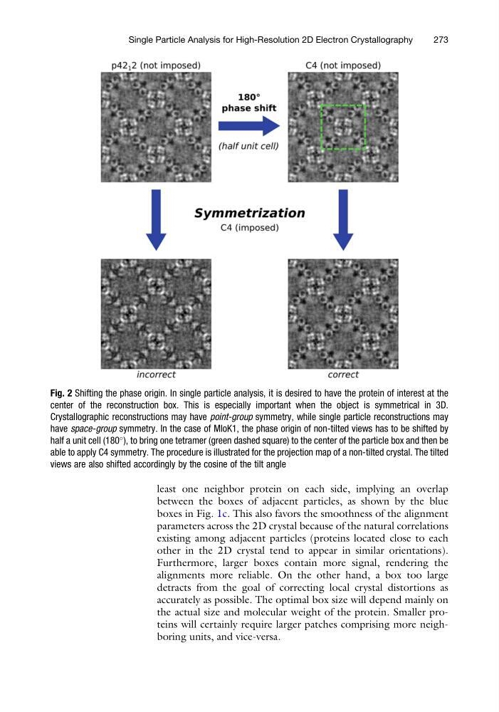
Fig. 2 Shifting the phase origin. In single particle analysis, it is desired to have the protein of interest at the
center of the reconstruction box. This is especially important when the object is symmetrical in 3D.
Crystallographic reconstructions may have point-group symmetry, while single particle reconstructions may
have space-group symmetry. In the case of MloK1, the phase origin of non-tilted views has to be shifted by
half a unit cell (180
), to bring one tetramer (green dashed square) to the center of the particle box and then be
able to apply C4 symmetry. The procedure is illustrated for the projection map of a non-tilted crystal. The tilted
views are also shifted accordingly by the cosine of the tilt angle
Single Particle Analysis for High-Resolution 2D Electron Crystallography
273
between the boxes of adjacent particles, as shown by the blue
boxes in Fig. 1c. This also favors the smoothness of the alignment
parameters across the 2D crystal because of the natural correlations
existing among adjacent particles (proteins located close to each
other in the 2D crystal tend to appear in similar orientations).
Furthermore, larger boxes contain more signal, rendering the
alignments more reliable. On the other hand, a box too large
detracts from the goal of correcting local crystal distortions as
accurately as possible. The optimal box size will depend mainly on
the actual size and molecular weight of the protein. Smaller proteins will certainly require larger patches comprising more neighboring units, and vice-versa.
Fig. 2 Shifting the phase origin. In single particle analysis, it is desired to have the protein of interest at the
center of the reconstruction box. This is especially important when the object is symmetrical in 3D.
Crystallographic reconstructions may have point-group symmetry, while single particle reconstructions may
have space-group symmetry. In the case of MloK1, the phase origin of non-tilted views has to be shifted by
half a unit cell (180
), to bring one tetramer (green dashed square) to the center of the particle box and then be
able to apply C4 symmetry. The procedure is illustrated for the projection map of a non-tilted crystal. The tilted
views are also shifted accordingly by the cosine of the tilt angle
Single Particle Analysis for High-Resolution 2D Electron Crystallography
273