

10. In the carbon-sandwich method, the size of the second carbon
film should not be too large. If the carbon film is too large, it
will fold to the other side of the grid and increase the thickness
of the grid specimen.
11. Spot size will need to be set to a step that can generate an
appropriate dose rate during the period of exposure time. One
should also consider about the size of the condenser aperture
in parallel when setting the spot size.
12. The camera length used for electron diffraction needs to be
determined by crystal quality and target resolution. An ideal
camera length provides a diffraction pattern with the highest
resolution reflection near the image edge.
Acknowledgments
We thank the Eyring Materials Center (EMC) at Arizona State
University (ASU) for providing the resources. Electron crystallographic data of the AQP0 and MloK1 2D crystals was the work in
Dr. Thomas Walz and Dr. Henning Stahlberg laboratories supported by the NIH grants, respectively.
References
1. Sonnhammer EL et al (1998) A hidden Markov model for predicting transmembrane helices in protein sequences. Proc Int Conf Intell
Syst Mol Biol 6:175–182
2. Wallin E, von Heijne G (1998) Genome-wide
analysis of integral membrane proteins from
eubacterial, archaean, and eukaryotic organisms. Protein Sci 7(4):1029–1038
3. Sachs JN, Engelman DM (2006) Introduction
to the membrane protein reviews: the interplay
of structure, dynamics, and environment in
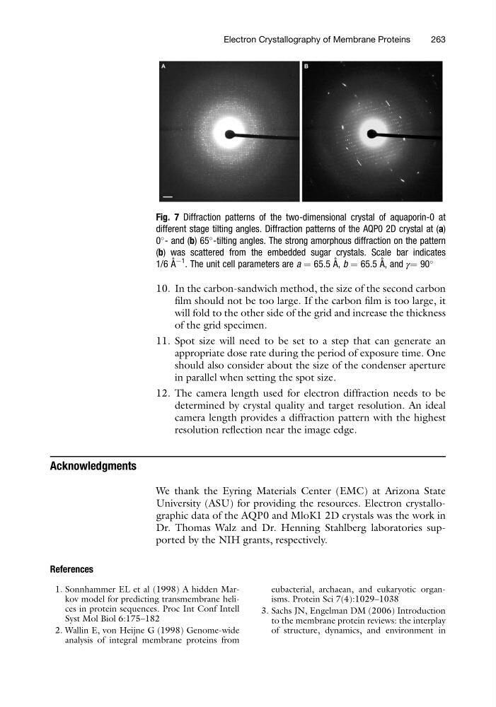
Fig. 7 Diffraction patterns of the two-dimensional crystal of aquaporin-0 at
different stage tilting angles. Diffraction patterns of the AQP0 2D crystal at (a)
0
- and (b) 65
-tilting angles. The strong amorphous diffraction on the pattern
(b) was scattered from the embedded sugar crystals. Scale bar indicates
1/6 A ˚ À1 . The unit cell parameters are a ¼ 65.5 A ˚ , b ¼ 65.5 A ˚ , and γ¼ 90
Electron Crystallography of Membrane Proteins
263
film should not be too large. If the carbon film is too large, it
will fold to the other side of the grid and increase the thickness
of the grid specimen.
11. Spot size will need to be set to a step that can generate an
appropriate dose rate during the period of exposure time. One
should also consider about the size of the condenser aperture
in parallel when setting the spot size.
12. The camera length used for electron diffraction needs to be
determined by crystal quality and target resolution. An ideal
camera length provides a diffraction pattern with the highest
resolution reflection near the image edge.
Acknowledgments
We thank the Eyring Materials Center (EMC) at Arizona State
University (ASU) for providing the resources. Electron crystallographic data of the AQP0 and MloK1 2D crystals was the work in
Dr. Thomas Walz and Dr. Henning Stahlberg laboratories supported by the NIH grants, respectively.
References
1. Sonnhammer EL et al (1998) A hidden Markov model for predicting transmembrane helices in protein sequences. Proc Int Conf Intell
Syst Mol Biol 6:175–182
2. Wallin E, von Heijne G (1998) Genome-wide
analysis of integral membrane proteins from
eubacterial, archaean, and eukaryotic organisms. Protein Sci 7(4):1029–1038
3. Sachs JN, Engelman DM (2006) Introduction
to the membrane protein reviews: the interplay
of structure, dynamics, and environment in
Fig. 7 Diffraction patterns of the two-dimensional crystal of aquaporin-0 at
different stage tilting angles. Diffraction patterns of the AQP0 2D crystal at (a)
0
- and (b) 65
-tilting angles. The strong amorphous diffraction on the pattern
(b) was scattered from the embedded sugar crystals. Scale bar indicates
1/6 A ˚ À1 . The unit cell parameters are a ¼ 65.5 A ˚ , b ¼ 65.5 A ˚ , and γ¼ 90
Electron Crystallography of Membrane Proteins
263