

the two carbon films sandwich the crystal specimen in the middle,
and this provides additional protection and keep the crystals
hydrated while imaging in the high vacuum. However, the downside of the carbon-sandwich method is that the specimen thickness
is not easy to control. The specimen thickness may be affected by
the variation of humidity, sugar concentration, and the thickness of
carbon film used.
Once a sugar-embedded 2D crystal is frozen, it will be transferred onto the TEM specimen stage for data collection. The
process of the cryo-specimen transfer can be accomplished using a
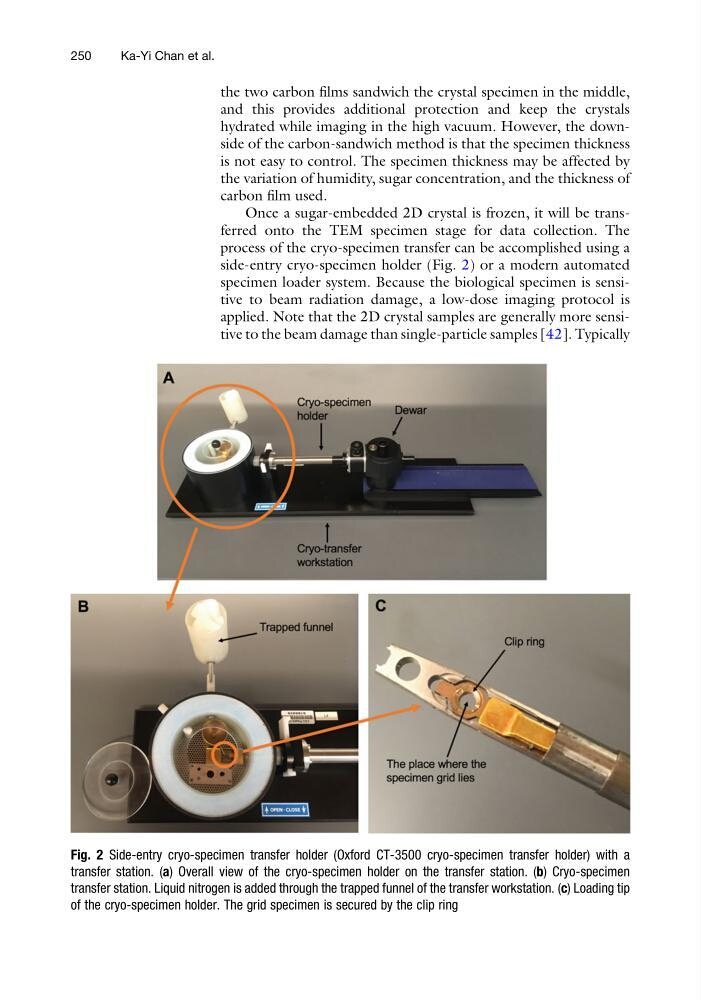
side-entry cryo-specimen holder (Fig. 2) or a modern automated
specimen loader system. Because the biological specimen is sensitive to beam radiation damage, a low-dose imaging protocol is
applied. Note that the 2D crystal samples are generally more sensitive to the beam damage than single-particle samples [42]. Typically
Fig. 2 Side-entry cryo-specimen transfer holder (Oxford CT-3500 cryo-specimen transfer holder) with a
transfer station. (a) Overall view of the cryo-specimen holder on the transfer station. (b) Cryo-specimen
transfer station. Liquid nitrogen is added through the trapped funnel of the transfer workstation. (c) Loading tip
of the cryo-specimen holder. The grid specimen is secured by the clip ring
250
Ka-Yi Chan et al.
and this provides additional protection and keep the crystals
hydrated while imaging in the high vacuum. However, the downside of the carbon-sandwich method is that the specimen thickness
is not easy to control. The specimen thickness may be affected by
the variation of humidity, sugar concentration, and the thickness of
carbon film used.
Once a sugar-embedded 2D crystal is frozen, it will be transferred onto the TEM specimen stage for data collection. The
process of the cryo-specimen transfer can be accomplished using a
side-entry cryo-specimen holder (Fig. 2) or a modern automated
specimen loader system. Because the biological specimen is sensitive to beam radiation damage, a low-dose imaging protocol is
applied. Note that the 2D crystal samples are generally more sensitive to the beam damage than single-particle samples [42]. Typically
Fig. 2 Side-entry cryo-specimen transfer holder (Oxford CT-3500 cryo-specimen transfer holder) with a
transfer station. (a) Overall view of the cryo-specimen holder on the transfer station. (b) Cryo-specimen
transfer station. Liquid nitrogen is added through the trapped funnel of the transfer workstation. (c) Loading tip
of the cryo-specimen holder. The grid specimen is secured by the clip ring
250
Ka-Yi Chan et al.