

7. From here, the grid is blotted as in Subheading 3.3.1 above, by
being placed on the two layers of Whatman filter paper without
releasing it from the forceps.
8. The grid is picked up, air-dried for 10–15 s, and hand-plunged
into liquid nitrogen, either for immediate or later use (see Note
30).
3.3.3 Initial Assessment
of Cryo-EM Conditions
Prior to data collection, it is often necessary to refine sample
preparation conditions after an initial assessment by cryo-EM.
Vitrified samples stored in liquid nitrogen are transferred into a
cryogenic transmission electron microscope and evaluated for ice
thickness and crystal integrity. If ice is too thick (i.e., the beam does
not penetrate the sample), it may be necessary to blot for longer
periods of time, or to alter sample buffer components. If the ice is
too thin, sample will be dehydrated and freeze-dried, rather than
vitrified, resulting in loss of crystalline order, and shorter blot times
and/or buffer alterations may be warranted. If ice thickness is ideal
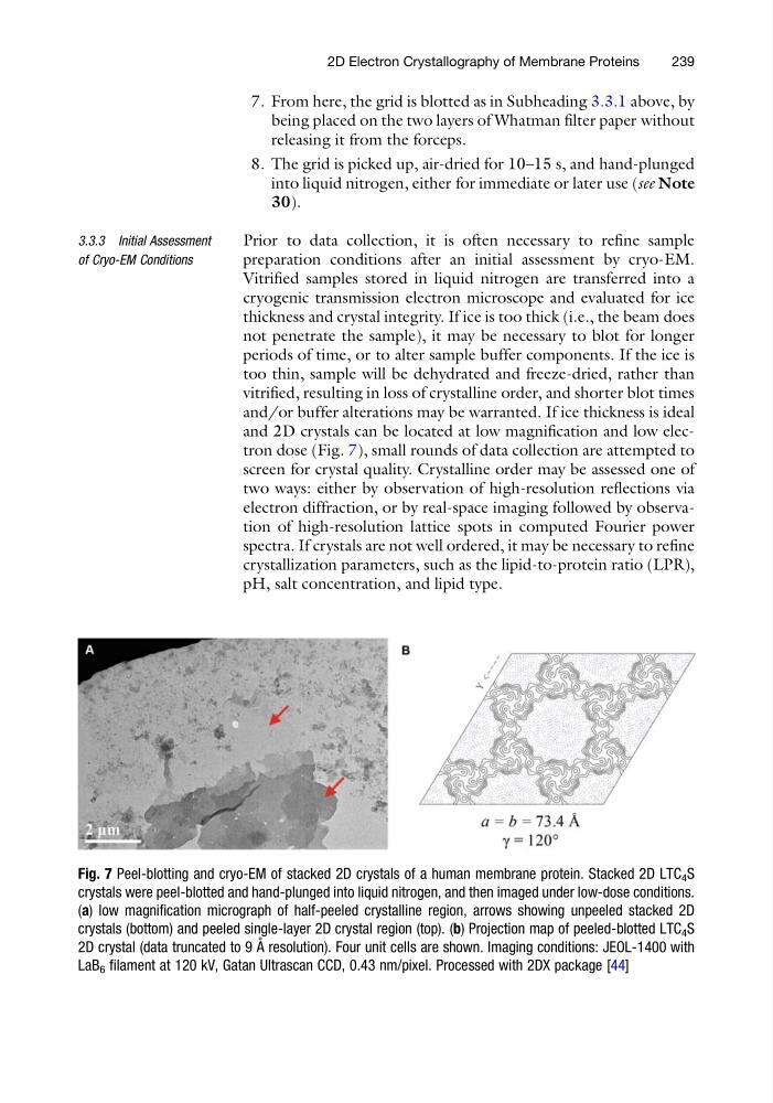
and 2D crystals can be located at low magnification and low electron dose (Fig. 7), small rounds of data collection are attempted to
screen for crystal quality. Crystalline order may be assessed one of
two ways: either by observation of high-resolution reflections via
electron diffraction, or by real-space imaging followed by observation of high-resolution lattice spots in computed Fourier power
spectra. If crystals are not well ordered, it may be necessary to refine
crystallization parameters, such as the lipid-to-protein ratio (LPR),
pH, salt concentration, and lipid type.
Fig. 7 Peel-blotting and cryo-EM of stacked 2D crystals of a human membrane protein. Stacked 2D LTC 4 S
crystals were peel-blotted and hand-plunged into liquid nitrogen, and then imaged under low-dose conditions.
(a) low magnification micrograph of half-peeled crystalline region, arrows showing unpeeled stacked 2D
crystals (bottom) and peeled single-layer 2D crystal region (top). (b) Projection map of peeled-blotted LTC 4 S
2D crystal (data truncated to 9 A ˚ resolution). Four unit cells are shown. Imaging conditions: JEOL-1400 with
LaB 6 filament at 120 kV, Gatan Ultrascan CCD, 0.43 nm/pixel. Processed with 2DX package [44]
2D Electron Crystallography of Membrane Proteins
239
being placed on the two layers of Whatman filter paper without
releasing it from the forceps.
8. The grid is picked up, air-dried for 10–15 s, and hand-plunged
into liquid nitrogen, either for immediate or later use (see Note
30).
3.3.3 Initial Assessment
of Cryo-EM Conditions
Prior to data collection, it is often necessary to refine sample
preparation conditions after an initial assessment by cryo-EM.
Vitrified samples stored in liquid nitrogen are transferred into a
cryogenic transmission electron microscope and evaluated for ice
thickness and crystal integrity. If ice is too thick (i.e., the beam does
not penetrate the sample), it may be necessary to blot for longer
periods of time, or to alter sample buffer components. If the ice is
too thin, sample will be dehydrated and freeze-dried, rather than
vitrified, resulting in loss of crystalline order, and shorter blot times
and/or buffer alterations may be warranted. If ice thickness is ideal
and 2D crystals can be located at low magnification and low electron dose (Fig. 7), small rounds of data collection are attempted to
screen for crystal quality. Crystalline order may be assessed one of
two ways: either by observation of high-resolution reflections via
electron diffraction, or by real-space imaging followed by observation of high-resolution lattice spots in computed Fourier power
spectra. If crystals are not well ordered, it may be necessary to refine
crystallization parameters, such as the lipid-to-protein ratio (LPR),
pH, salt concentration, and lipid type.
Fig. 7 Peel-blotting and cryo-EM of stacked 2D crystals of a human membrane protein. Stacked 2D LTC 4 S
crystals were peel-blotted and hand-plunged into liquid nitrogen, and then imaged under low-dose conditions.
(a) low magnification micrograph of half-peeled crystalline region, arrows showing unpeeled stacked 2D
crystals (bottom) and peeled single-layer 2D crystal region (top). (b) Projection map of peeled-blotted LTC 4 S
2D crystal (data truncated to 9 A ˚ resolution). Four unit cells are shown. Imaging conditions: JEOL-1400 with
LaB 6 filament at 120 kV, Gatan Ultrascan CCD, 0.43 nm/pixel. Processed with 2DX package [44]
2D Electron Crystallography of Membrane Proteins
239