

4. Ensure that dose-fractionation is enabled for record images and
the frames are saved to a specific directory with tilt angle and
navigator position appended to the file name.
5. For each tilt series collected, you should obtain an filename.
mrc, filename.mrc.mdoc, and filename.log file as well as the
individual dose-fractionated images which are saved to a separate directory.
3.4 Tomogram
Generation
Tomogram generation is performed using Etomo in IMOD. A
tutorial for reconstructing tomograms with Etomo is available on
the IMOD website (https://bio3d.colorado.edu/imod/doc/
etomoTutorial.html). In this section, we will mainly focus on the
modification to the tutorial for the processing of the dataset
obtained in Subheading 3.3. An example tomogram is shown in
Fig. 3.
1. Move each tilt series filename.mrc together with respective
filename.mrc.mdoc, and filename.log files into individual directories/folders on your processing workstation.
2. For morphological studies, filename.mrc tilt series file from
SerialEM should be sufficient. For subtomogram averaging,
the dose-fractionated frames should be aligned with MotionCor2 and assembled into a filename.mrc tilt series stack. This
can be done by using the “newstack” function in IMOD.
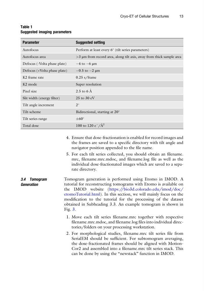
Table 1
Suggested imaging parameters
Parameter
Suggested setting
Autofocus
Perform at least every 6
(tilt series parameters)
Autofocus area
>3 μm from record area, along tilt axis, away from thick sample area
Defocus (-Volta phase plate)
À4 to À6 μm
Defocus (+Volta phase plate)
À0.5 to À2 μm
K2 frame rate
0.25 s/frame
K2 mode
Super resolution
Pixel size
2.5 to 6 A ˚
Slit width (energy filter)
25 to 30 eV
Tilt angle increment
2
Tilt-scheme
Bidirectional, starting at 20
Tilt series range
Æ60
Total dose
100 to 120 e
À
/A ˚ 2
Cryo-ET of Cellular Structures
13
the frames are saved to a specific directory with tilt angle and
navigator position appended to the file name.
5. For each tilt series collected, you should obtain an filename.
mrc, filename.mrc.mdoc, and filename.log file as well as the
individual dose-fractionated images which are saved to a separate directory.
3.4 Tomogram
Generation
Tomogram generation is performed using Etomo in IMOD. A
tutorial for reconstructing tomograms with Etomo is available on
the IMOD website (https://bio3d.colorado.edu/imod/doc/
etomoTutorial.html). In this section, we will mainly focus on the
modification to the tutorial for the processing of the dataset
obtained in Subheading 3.3. An example tomogram is shown in
Fig. 3.
1. Move each tilt series filename.mrc together with respective
filename.mrc.mdoc, and filename.log files into individual directories/folders on your processing workstation.
2. For morphological studies, filename.mrc tilt series file from
SerialEM should be sufficient. For subtomogram averaging,
the dose-fractionated frames should be aligned with MotionCor2 and assembled into a filename.mrc tilt series stack. This
can be done by using the “newstack” function in IMOD.
Table 1
Suggested imaging parameters
Parameter
Suggested setting
Autofocus
Perform at least every 6
(tilt series parameters)
Autofocus area
>3 μm from record area, along tilt axis, away from thick sample area
Defocus (-Volta phase plate)
À4 to À6 μm
Defocus (+Volta phase plate)
À0.5 to À2 μm
K2 frame rate
0.25 s/frame
K2 mode
Super resolution
Pixel size
2.5 to 6 A ˚
Slit width (energy filter)
25 to 30 eV
Tilt angle increment
2
Tilt-scheme
Bidirectional, starting at 20
Tilt series range
Æ60
Total dose
100 to 120 e
À
/A ˚ 2
Cryo-ET of Cellular Structures
13