

changing z-height. Tilt to 0
and recenter screen using x/y
controls. Repeat until change in position of detectable feature
is minimal.
5. Collect montage of whole grid (software path: Navigator !
Montaging & Grids ! Setup Full Montage). Ensure image
overlap is set to 20%.
3.3.2 Collection
of Polygon Maps
1. Switch magnification and setup low dose (see Note 35). Suggested mode magnifications are: Record/focus/preview
0.3 nm/pixel, View ~3 nm/pixel.
2. Align whole grid montage to view mode (see Note 35).
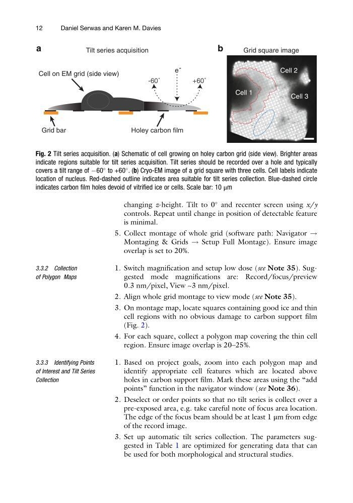
3. On montage map, locate squares containing good ice and thin
cell regions with no obvious damage to carbon support film
(Fig. 2).
4. For each square, collect a polygon map covering the thin cell
region. Ensure image overlap is 20–25%.
3.3.3 Identifying Points
of Interest and Tilt Series
Collection
1. Based on project goals, zoom into each polygon map and
identify appropriate cell features which are located above
holes in carbon support film. Mark these areas using the “add
points” function in the navigator window (see Note 36).
2. Deselect or order points so that no tilt series is collect over a
pre-exposed area, e.g. take careful note of focus area location.
The edge of the focus beam should be at least 1 μm from edge
of the record image.
3. Set up automatic tilt series collection. The parameters suggested in Table 1 are optimized for generating data that can
be used for both morphological and structural studies.
a
Tilt series acquisition
b
e -
-60˚
+60˚
Cell on EM grid (side view)
Holey carbon film
Grid bar
Cell 1
Cell 2
Cell 3
Grid square image
Fig. 2 Tilt series acquisition. (a) Schematic of cell growing on holey carbon grid (side view). Brighter areas
indicate regions suitable for tilt series acquisition. Tilt series should be recorded over a hole and typically
covers a tilt range of À60
to +60
. (b) Cryo-EM image of a grid square with three cells. Cell labels indicate
location of nucleus. Red-dashed outline indicates area suitable for tilt series collection. Blue-dashed circle
indicates carbon film holes devoid of vitrified ice or cells. Scale bar: 10 μm
12
Daniel Serwas and Karen M. Davies
and recenter screen using x/y
controls. Repeat until change in position of detectable feature
is minimal.
5. Collect montage of whole grid (software path: Navigator !
Montaging & Grids ! Setup Full Montage). Ensure image
overlap is set to 20%.
3.3.2 Collection
of Polygon Maps
1. Switch magnification and setup low dose (see Note 35). Suggested mode magnifications are: Record/focus/preview
0.3 nm/pixel, View ~3 nm/pixel.
2. Align whole grid montage to view mode (see Note 35).
3. On montage map, locate squares containing good ice and thin
cell regions with no obvious damage to carbon support film
(Fig. 2).
4. For each square, collect a polygon map covering the thin cell
region. Ensure image overlap is 20–25%.
3.3.3 Identifying Points
of Interest and Tilt Series
Collection
1. Based on project goals, zoom into each polygon map and
identify appropriate cell features which are located above
holes in carbon support film. Mark these areas using the “add
points” function in the navigator window (see Note 36).
2. Deselect or order points so that no tilt series is collect over a
pre-exposed area, e.g. take careful note of focus area location.
The edge of the focus beam should be at least 1 μm from edge
of the record image.
3. Set up automatic tilt series collection. The parameters suggested in Table 1 are optimized for generating data that can
be used for both morphological and structural studies.
a
Tilt series acquisition
b
e -
-60˚
+60˚
Cell on EM grid (side view)
Holey carbon film
Grid bar
Cell 1
Cell 2
Cell 3
Grid square image
Fig. 2 Tilt series acquisition. (a) Schematic of cell growing on holey carbon grid (side view). Brighter areas
indicate regions suitable for tilt series acquisition. Tilt series should be recorded over a hole and typically
covers a tilt range of À60
to +60
. (b) Cryo-EM image of a grid square with three cells. Cell labels indicate
location of nucleus. Red-dashed outline indicates area suitable for tilt series collection. Blue-dashed circle
indicates carbon film holes devoid of vitrified ice or cells. Scale bar: 10 μm
12
Daniel Serwas and Karen M. Davies