

readily obtained or purified, negative-stain can not only significantly lower the barrier to a well-designed CryoEM experiment
but can sometimes also serve to add biological information in cases
when CryoEM is not viable [8].
While there are unique elements to screening in CryoEM,
including minimizing grid contamination [9] and getting particles
distributed in different orientations in thin layers of vitreous ice
[10], some elements are common and can be quickly accessed using
negative-stain. Additionally, negative-stain can afford tremendous
benefits and advantages as both a prior and complementary tool for
CryoEM, including:
The viability, stability, purity, and homogeneity of particles can be
quickly accessed [6]; Samples can be stained within seconds after
preparations are made and therefore can spend little time outside of
their optimal biochemical conditions; Concentrations can be checked
and an initial starting point for CryoEM determined (see Note 1);
Greater contrast of the sample when compared to CryoEM allowing
for smaller proteins to be imaged [11]; Initial datasets can be
obtained for single particle averaging and electron crystallography
and result in maps upwards of 14–20 A ˚ in resolution allowing quick
assessment [12]; Buffers, salts, and other conditions can be optimized;
Cheaper than a CryoEM experiment; Once the grid is stained it can
be left at ambient temperatures for years either for archiving or while
waiting for microscope time; Grids can be checked using the majority
of electron microscopes already available at most institutions (including those without direct electron detectors); Imaging and interpretation can rely less on perfect alignment of the beam; Grids can
withstand more mechanical bending than in CryoEM; Technique is
more accessible to non-experts and provides a good gateway to CryoEM
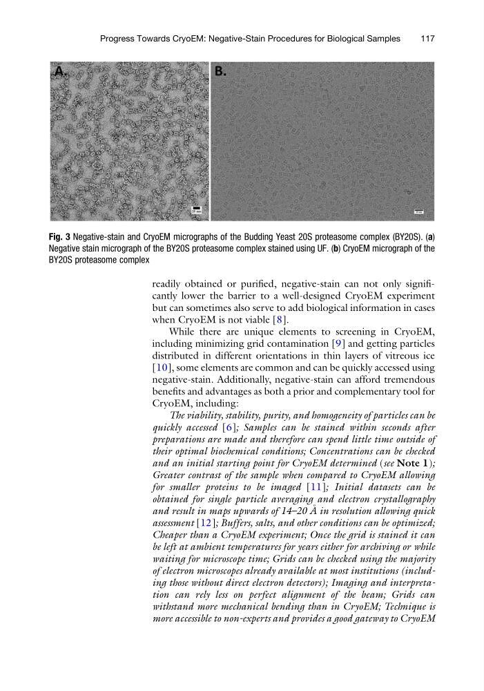
Fig. 3 Negative-stain and CryoEM micrographs of the Budding Yeast 20S proteasome complex (BY20S). (a)
Negative stain micrograph of the BY20S proteasome complex stained using UF. (b) CryoEM micrograph of the
BY20S proteasome complex
Progress Towards CryoEM: Negative-Stain Procedures for Biological Samples
117
but can sometimes also serve to add biological information in cases
when CryoEM is not viable [8].
While there are unique elements to screening in CryoEM,
including minimizing grid contamination [9] and getting particles
distributed in different orientations in thin layers of vitreous ice
[10], some elements are common and can be quickly accessed using
negative-stain. Additionally, negative-stain can afford tremendous
benefits and advantages as both a prior and complementary tool for
CryoEM, including:
The viability, stability, purity, and homogeneity of particles can be
quickly accessed [6]; Samples can be stained within seconds after
preparations are made and therefore can spend little time outside of
their optimal biochemical conditions; Concentrations can be checked
and an initial starting point for CryoEM determined (see Note 1);
Greater contrast of the sample when compared to CryoEM allowing
for smaller proteins to be imaged [11]; Initial datasets can be
obtained for single particle averaging and electron crystallography
and result in maps upwards of 14–20 A ˚ in resolution allowing quick
assessment [12]; Buffers, salts, and other conditions can be optimized;
Cheaper than a CryoEM experiment; Once the grid is stained it can
be left at ambient temperatures for years either for archiving or while
waiting for microscope time; Grids can be checked using the majority
of electron microscopes already available at most institutions (including those without direct electron detectors); Imaging and interpretation can rely less on perfect alignment of the beam; Grids can
withstand more mechanical bending than in CryoEM; Technique is
more accessible to non-experts and provides a good gateway to CryoEM
Fig. 3 Negative-stain and CryoEM micrographs of the Budding Yeast 20S proteasome complex (BY20S). (a)
Negative stain micrograph of the BY20S proteasome complex stained using UF. (b) CryoEM micrograph of the
BY20S proteasome complex
Progress Towards CryoEM: Negative-Stain Procedures for Biological Samples
117