

description of strata, a diversity of microbial functional groups alternate within a few
mm of depth, adapting to steep physicochemical gradients (in light penetration, O 2
and H 2 S; Fig. 3.2a) and exploiting every possible ecological niche generated within
the mat (van Gemerden 1993). The same taxa (even species) are often found in
similar arrangements and niches, which is an indication of the cosmopolitanism of
these microbes.
It is very important to bear in mind that these gradients vary on a diel basis, with
O 2 supersaturation during daytime when photoautotrophs are active and accumulation of H 2 S at night, when oxygenic photosynthesis is halted. Therefore, the O 2 /H 2 S
interface schematized in Fig. 3.2a is highly dynamic and critical to the functioning of
the microbial mat consortium. Furthermore, a rather radical hypothesis has proposed
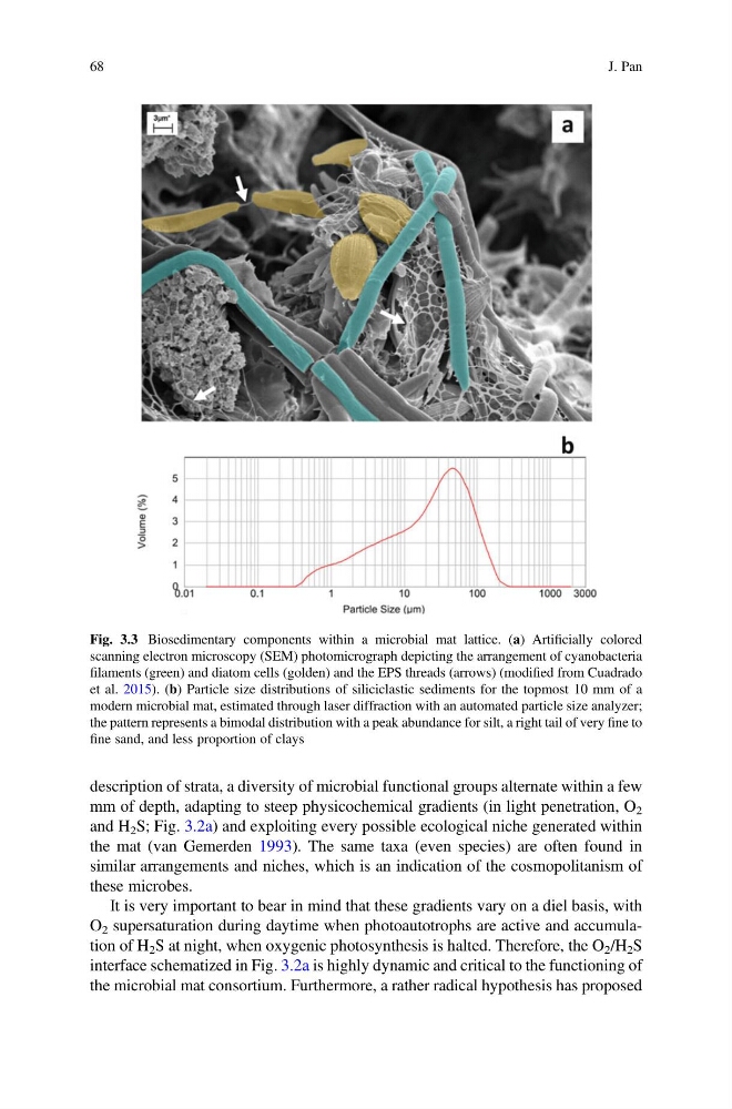
Fig. 3.3 Biosedimentary components within a microbial mat lattice. (a) Artificially colored
scanning electron microscopy (SEM) photomicrograph depicting the arrangement of cyanobacteria
filaments (green) and diatom cells (golden) and the EPS threads (arrows) (modified from Cuadrado
et al. 2015). (b) Particle size distributions of siliciclastic sediments for the topmost 10 mm of a
modern microbial mat, estimated through laser diffraction with an automated particle size analyzer;
the pattern represents a bimodal distribution with a peak abundance for silt, a right tail of very fine to
fine sand, and less proportion of clays
68
J. Pan
mm of depth, adapting to steep physicochemical gradients (in light penetration, O 2
and H 2 S; Fig. 3.2a) and exploiting every possible ecological niche generated within
the mat (van Gemerden 1993). The same taxa (even species) are often found in
similar arrangements and niches, which is an indication of the cosmopolitanism of
these microbes.
It is very important to bear in mind that these gradients vary on a diel basis, with
O 2 supersaturation during daytime when photoautotrophs are active and accumulation of H 2 S at night, when oxygenic photosynthesis is halted. Therefore, the O 2 /H 2 S
interface schematized in Fig. 3.2a is highly dynamic and critical to the functioning of
the microbial mat consortium. Furthermore, a rather radical hypothesis has proposed
Fig. 3.3 Biosedimentary components within a microbial mat lattice. (a) Artificially colored
scanning electron microscopy (SEM) photomicrograph depicting the arrangement of cyanobacteria
filaments (green) and diatom cells (golden) and the EPS threads (arrows) (modified from Cuadrado
et al. 2015). (b) Particle size distributions of siliciclastic sediments for the topmost 10 mm of a
modern microbial mat, estimated through laser diffraction with an automated particle size analyzer;
the pattern represents a bimodal distribution with a peak abundance for silt, a right tail of very fine to
fine sand, and less proportion of clays
68
J. Pan