

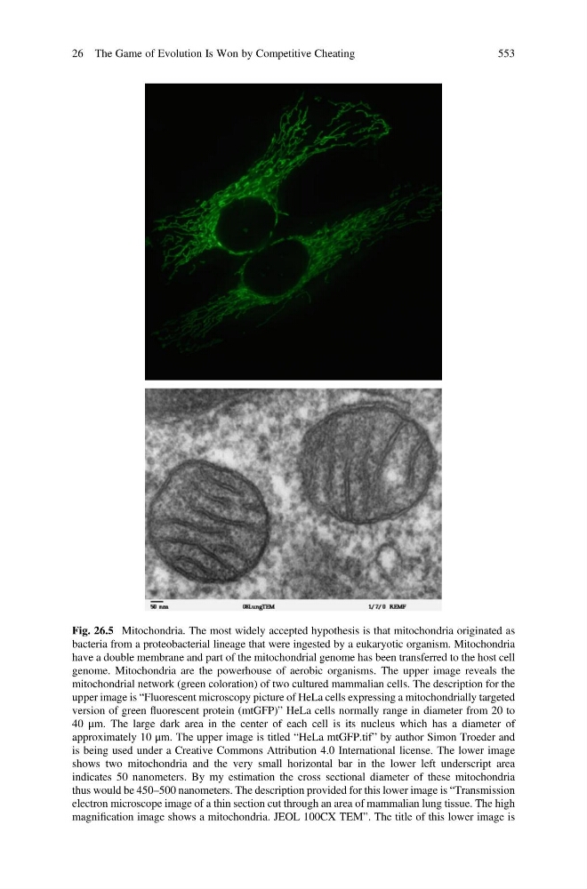
Fig. 26.5 Mitochondria. The most widely accepted hypothesis is that mitochondria originated as
bacteria from a proteobacterial lineage that were ingested by a eukaryotic organism. Mitochondria
have a double membrane and part of the mitochondrial genome has been transferred to the host cell
genome. Mitochondria are the powerhouse of aerobic organisms. The upper image reveals the
mitochondrial network (green coloration) of two cultured mammalian cells. The description for the
upper image is “Fluorescent microscopy picture of HeLa cells expressing a mitochondrially targeted
version of green fluorescent protein (mtGFP)” HeLa cells normally range in diameter from 20 to
40 μm. The large dark area in the center of each cell is its nucleus which has a diameter of
approximately 10 μm. The upper image is titled “HeLa mtGFP.tif” by author Simon Troeder and
is being used under a Creative Commons Attribution 4.0 International license. The lower image
shows two mitochondria and the very small horizontal bar in the lower left underscript area
indicates 50 nanometers. By my estimation the cross sectional diameter of these mitochondria
thus would be 450–500 nanometers. The description provided for this lower image is “Transmission
electron microscope image of a thin section cut through an area of mammalian lung tissue. The high
magnification image shows a mitochondria. JEOL 100CX TEM”. The title of this lower image is
26 The Game of Evolution Is Won by Competitive Cheating
553
bacteria from a proteobacterial lineage that were ingested by a eukaryotic organism. Mitochondria
have a double membrane and part of the mitochondrial genome has been transferred to the host cell
genome. Mitochondria are the powerhouse of aerobic organisms. The upper image reveals the
mitochondrial network (green coloration) of two cultured mammalian cells. The description for the
upper image is “Fluorescent microscopy picture of HeLa cells expressing a mitochondrially targeted
version of green fluorescent protein (mtGFP)” HeLa cells normally range in diameter from 20 to
40 μm. The large dark area in the center of each cell is its nucleus which has a diameter of
approximately 10 μm. The upper image is titled “HeLa mtGFP.tif” by author Simon Troeder and
is being used under a Creative Commons Attribution 4.0 International license. The lower image
shows two mitochondria and the very small horizontal bar in the lower left underscript area
indicates 50 nanometers. By my estimation the cross sectional diameter of these mitochondria
thus would be 450–500 nanometers. The description provided for this lower image is “Transmission
electron microscope image of a thin section cut through an area of mammalian lung tissue. The high
magnification image shows a mitochondria. JEOL 100CX TEM”. The title of this lower image is
26 The Game of Evolution Is Won by Competitive Cheating
553