

“loops”). A specific dimeric structure has been chosen to represent
each class as a practical example and a table summarizing the
average values of the descriptors provided, together with confidence tests. General comments are also added in the conclusion
section.
2 Methods
2.1 Preparing
and Analyzing the PDB
Files of Homodimers
Since the crystallographic structure might not correspond to the
effective in vivo/ex vivo tri-dimensional assembly, a careful inspection of the PDB file is recommended.
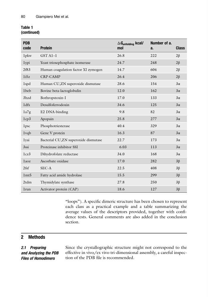
Table 1
(continued)
PDB
code
Protein
ΔG unfolding kcal/
mol
Number of a.
a.
Class
1pkw
GST A1–1
26.8
222
2β
1ypi
Yeast triosephosphate isomerase
24.7
248
2β
2f83
Human coagulation factor XI zymogen
14.7
604
2β
1i5z
CRP-CAMP
26.4
206
2β
1spd
Human CU,ZN superoxide dismutase
28.6
154
3α
1beb
Bovine beta-lactoglobulin
12.0
162
3α
3hzd
Bothropstoxin-I
17.0
133
3α
1dfx
Desulfoferrodoxin
34.6
125
3α
1a7g
E2 DNA-binding
9.8
82
3α
1cp3
Apopain
25.8
277
3α
1psc
Phosphotriesterase
40.4
329
3α
1vqb
Gene V protein
16.3
87
3α
1yai
Bacterial CU,ZN superoxide dismutase
22.7
173
3α
3ssi
Proteinase inhibitor SSI
6.03
113
3α
1cz3
Dihydrofolate reductase
34.0
168
3α
1aoz
Ascorbate oxidase
17.0
282
3β
2fsf
SEC-A
22.5
408
3β
1mt5
Fatty acid amide hydrolase
15.5
299
3β
2tdm
Thymidylate synthase
27.8
250
3β
1run
Activator protein (CAP)
18.6
127
3β
80
Giampiero Mei et al.
each class as a practical example and a table summarizing the
average values of the descriptors provided, together with confidence tests. General comments are also added in the conclusion
section.
2 Methods
2.1 Preparing
and Analyzing the PDB
Files of Homodimers
Since the crystallographic structure might not correspond to the
effective in vivo/ex vivo tri-dimensional assembly, a careful inspection of the PDB file is recommended.
Table 1
(continued)
PDB
code
Protein
ΔG unfolding kcal/
mol
Number of a.
a.
Class
1pkw
GST A1–1
26.8
222
2β
1ypi
Yeast triosephosphate isomerase
24.7
248
2β
2f83
Human coagulation factor XI zymogen
14.7
604
2β
1i5z
CRP-CAMP
26.4
206
2β
1spd
Human CU,ZN superoxide dismutase
28.6
154
3α
1beb
Bovine beta-lactoglobulin
12.0
162
3α
3hzd
Bothropstoxin-I
17.0
133
3α
1dfx
Desulfoferrodoxin
34.6
125
3α
1a7g
E2 DNA-binding
9.8
82
3α
1cp3
Apopain
25.8
277
3α
1psc
Phosphotriesterase
40.4
329
3α
1vqb
Gene V protein
16.3
87
3α
1yai
Bacterial CU,ZN superoxide dismutase
22.7
173
3α
3ssi
Proteinase inhibitor SSI
6.03
113
3α
1cz3
Dihydrofolate reductase
34.0
168
3α
1aoz
Ascorbate oxidase
17.0
282
3β
2fsf
SEC-A
22.5
408
3β
1mt5
Fatty acid amide hydrolase
15.5
299
3β
2tdm
Thymidylate synthase
27.8
250
3β
1run
Activator protein (CAP)
18.6
127
3β
80
Giampiero Mei et al.