

N
0 -[(5
0 -phosphoribulosyl)formimino]-5-aminoimidazole-4-carboxamide-ribonucleotide binds, are the two tightly associated proteins constituting the IGPS allosteric enzyme, as shown in Fig. 4.
PRFAR effector binding accelerates glutamine hydrolysis by
ca. 5000-fold, with respect to the apo-IGPS enzyme [52].
As shown in Fig. 4, the outcome of the CNA method is a
coarse-grained picture of the division of the allosteric proteic system, which has a straightforward structural interpretation but it
also contains information on the communication flow within the
complex network of amino acid residues. In the case of IGPS, the
CNA method showed to be quite sensitive to the changes in
communication network induced by the allosteric regulator, allowing detection of suggested secondary structure elements and key
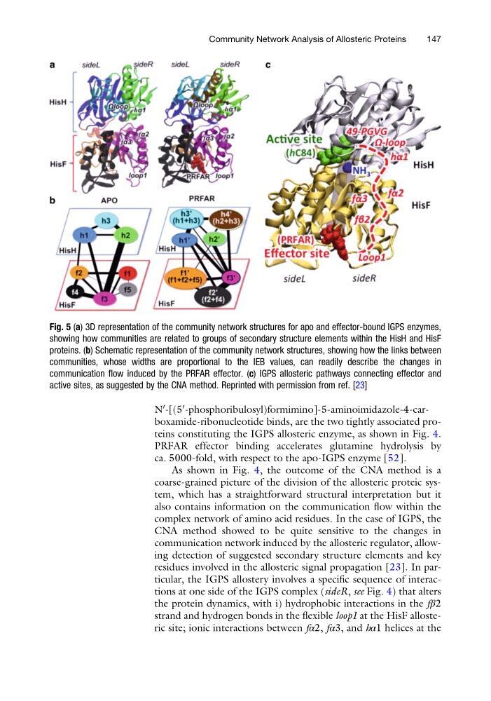
residues involved in the allosteric signal propagation [23]. In particular, the IGPS allostery involves a specific sequence of interactions at one side of the IGPS complex (sideR, see Fig. 4) that alters
the protein dynamics, with i) hydrophobic interactions in the fβ2
strand and hydrogen bonds in the flexible loop1 at the HisF allosteric site; ionic interactions between fα2, fα3, and hα1 helices at the
Fig. 5 (a) 3D representation of the community network structures for apo and effector-bound IGPS enzymes,
showing how communities are related to groups of secondary structure elements within the HisH and HisF
proteins. (b) Schematic representation of the community network structures, showing how the links between
communities, whose widths are proportional to the IEB values, can readily describe the changes in
communication flow induced by the PRFAR effector. (c) IGPS allosteric pathways connecting effector and
active sites, as suggested by the CNA method. Reprinted with permission from ref. [23]
Community Network Analysis of Allosteric Proteins
147
0 -[(5
0 -phosphoribulosyl)formimino]-5-aminoimidazole-4-carboxamide-ribonucleotide binds, are the two tightly associated proteins constituting the IGPS allosteric enzyme, as shown in Fig. 4.
PRFAR effector binding accelerates glutamine hydrolysis by
ca. 5000-fold, with respect to the apo-IGPS enzyme [52].
As shown in Fig. 4, the outcome of the CNA method is a
coarse-grained picture of the division of the allosteric proteic system, which has a straightforward structural interpretation but it
also contains information on the communication flow within the
complex network of amino acid residues. In the case of IGPS, the
CNA method showed to be quite sensitive to the changes in
communication network induced by the allosteric regulator, allowing detection of suggested secondary structure elements and key
residues involved in the allosteric signal propagation [23]. In particular, the IGPS allostery involves a specific sequence of interactions at one side of the IGPS complex (sideR, see Fig. 4) that alters
the protein dynamics, with i) hydrophobic interactions in the fβ2
strand and hydrogen bonds in the flexible loop1 at the HisF allosteric site; ionic interactions between fα2, fα3, and hα1 helices at the
Fig. 5 (a) 3D representation of the community network structures for apo and effector-bound IGPS enzymes,
showing how communities are related to groups of secondary structure elements within the HisH and HisF
proteins. (b) Schematic representation of the community network structures, showing how the links between
communities, whose widths are proportional to the IEB values, can readily describe the changes in
communication flow induced by the PRFAR effector. (c) IGPS allosteric pathways connecting effector and
active sites, as suggested by the CNA method. Reprinted with permission from ref. [23]
Community Network Analysis of Allosteric Proteins
147