

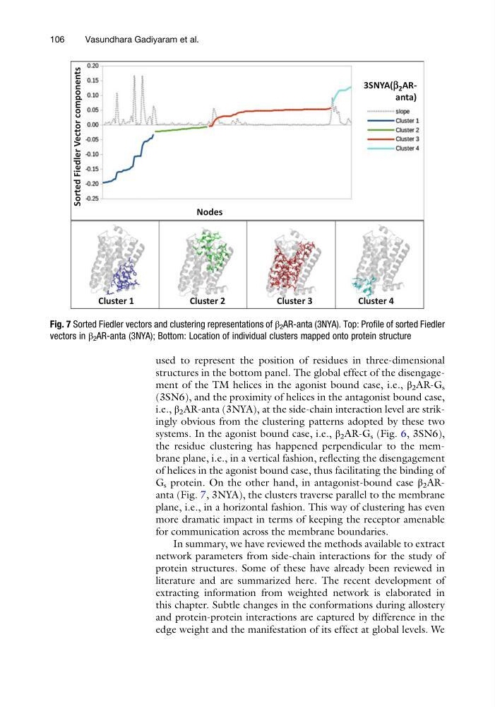
used to represent the position of residues in three-dimensional
structures in the bottom panel. The global effect of the disengagement of the TM helices in the agonist bound case, i.e., β 2 AR-G s
(3SN6), and the proximity of helices in the antagonist bound case,
i.e., β 2 AR-anta (3NYA), at the side-chain interaction level are strikingly obvious from the clustering patterns adopted by these two
systems. In the agonist bound case, i.e., β 2 AR-G s (Fig. 6, 3SN6),
the residue clustering has happened perpendicular to the membrane plane, i.e., in a vertical fashion, reflecting the disengagement
of helices in the agonist bound case, thus facilitating the binding of
G s protein. On the other hand, in antagonist-bound case β 2 ARanta (Fig. 7, 3NYA), the clusters traverse parallel to the membrane
plane, i.e., in a horizontal fashion. This way of clustering has even
more dramatic impact in terms of keeping the receptor amenable
for communication across the membrane boundaries.
In summary, we have reviewed the methods available to extract
network parameters from side-chain interactions for the study of
protein structures. Some of these have already been reviewed in
literature and are summarized here. The recent development of
extracting information from weighted network is elaborated in
this chapter. Subtle changes in the conformations during allostery
and protein-protein interactions are captured by difference in the
edge weight and the manifestation of its effect at global levels. We
Cluster 1
Cluster 2
Nodes
3SNYA(b 2 ARanta)
Sorted Fiedler Vector components
Cluster 3
Cluster 4
Fig. 7 Sorted Fiedler vectors and clustering representations of β 2 AR-anta (3NYA). Top: Profile of sorted Fiedler
vectors in β 2 AR-anta (3NYA); Bottom: Location of individual clusters mapped onto protein structure
106
Vasundhara Gadiyaram et al.
structures in the bottom panel. The global effect of the disengagement of the TM helices in the agonist bound case, i.e., β 2 AR-G s
(3SN6), and the proximity of helices in the antagonist bound case,
i.e., β 2 AR-anta (3NYA), at the side-chain interaction level are strikingly obvious from the clustering patterns adopted by these two
systems. In the agonist bound case, i.e., β 2 AR-G s (Fig. 6, 3SN6),
the residue clustering has happened perpendicular to the membrane plane, i.e., in a vertical fashion, reflecting the disengagement
of helices in the agonist bound case, thus facilitating the binding of
G s protein. On the other hand, in antagonist-bound case β 2 ARanta (Fig. 7, 3NYA), the clusters traverse parallel to the membrane
plane, i.e., in a horizontal fashion. This way of clustering has even
more dramatic impact in terms of keeping the receptor amenable
for communication across the membrane boundaries.
In summary, we have reviewed the methods available to extract
network parameters from side-chain interactions for the study of
protein structures. Some of these have already been reviewed in
literature and are summarized here. The recent development of
extracting information from weighted network is elaborated in
this chapter. Subtle changes in the conformations during allostery
and protein-protein interactions are captured by difference in the
edge weight and the manifestation of its effect at global levels. We
Cluster 1
Cluster 2
Nodes
3SNYA(b 2 ARanta)
Sorted Fiedler Vector components
Cluster 3
Cluster 4
Fig. 7 Sorted Fiedler vectors and clustering representations of β 2 AR-anta (3NYA). Top: Profile of sorted Fiedler
vectors in β 2 AR-anta (3NYA); Bottom: Location of individual clusters mapped onto protein structure
106
Vasundhara Gadiyaram et al.