

G-protein. Hence, it is interesting to note how changes in ligand
binding pocket translate into structural alterations in TM5, TM6
and TM7 which facilitate the binding of G s . Agonist interactions
with Ser203
5.42 and Ser207
5.46 give rise to a 2 A ˚ inward motion of
TM5 at Ser207
5.46 and 1.4 A ˚ inward movement at Pro211
5.50
[38]. This pull disturbs a highly conserved network of contacts
between Pro211
5.50
, Ile121
3.40
, Phe282
6.44 and Asn318
7.45
. As a
result, Ile121
3.40 and Phe282
6.44 are repositioned, along with a
rotation about Phe282
6.44
, leading to an outward displacement of
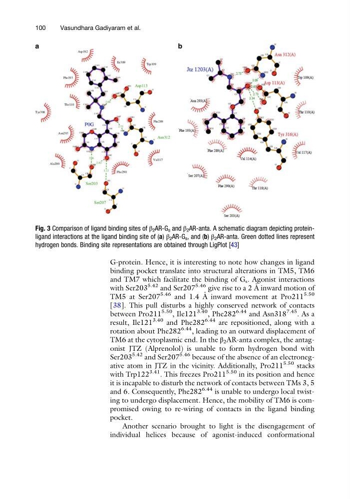
TM6 at the cytoplasmic end. In the β 2 AR-anta complex, the antagonist JTZ (Alprenolol) is unable to form hydrogen bond with
Ser203
5.42 and Ser207
5.46 because of the absence of an electronegative atom in JTZ in the vicinity. Additionally, Pro211
5.50 stacks
with Trp122
3.41
. This freezes Pro211
5.50 in its position and hence
it is incapable to disturb the network of contacts between TMs 3, 5
and 6. Consequently, Phe282
6.44 is unable to undergo local twisting to undergo displacement. Hence, the mobility of TM6 is compromised owing to re-wiring of contacts in the ligand binding
pocket.
Another scenario brought to light is the disengagement of
individual helices because of agonist-induced conformational
Fig. 3 Comparison of ligand binding sites of β 2 AR-G s and β 2 AR-anta. A schematic diagram depicting proteinligand interactions at the ligand binding site of (a) β 2 AR-G s , and (b) β 2 AR-anta. Green dotted lines represent
hydrogen bonds. Binding site representations are obtained through LigPlot [43]
100
Vasundhara Gadiyaram et al.
binding pocket translate into structural alterations in TM5, TM6
and TM7 which facilitate the binding of G s . Agonist interactions
with Ser203
5.42 and Ser207
5.46 give rise to a 2 A ˚ inward motion of
TM5 at Ser207
5.46 and 1.4 A ˚ inward movement at Pro211
5.50
[38]. This pull disturbs a highly conserved network of contacts
between Pro211
5.50
, Ile121
3.40
, Phe282
6.44 and Asn318
7.45
. As a
result, Ile121
3.40 and Phe282
6.44 are repositioned, along with a
rotation about Phe282
6.44
, leading to an outward displacement of
TM6 at the cytoplasmic end. In the β 2 AR-anta complex, the antagonist JTZ (Alprenolol) is unable to form hydrogen bond with
Ser203
5.42 and Ser207
5.46 because of the absence of an electronegative atom in JTZ in the vicinity. Additionally, Pro211
5.50 stacks
with Trp122
3.41
. This freezes Pro211
5.50 in its position and hence
it is incapable to disturb the network of contacts between TMs 3, 5
and 6. Consequently, Phe282
6.44 is unable to undergo local twisting to undergo displacement. Hence, the mobility of TM6 is compromised owing to re-wiring of contacts in the ligand binding
pocket.
Another scenario brought to light is the disengagement of
individual helices because of agonist-induced conformational
Fig. 3 Comparison of ligand binding sites of β 2 AR-G s and β 2 AR-anta. A schematic diagram depicting proteinligand interactions at the ligand binding site of (a) β 2 AR-G s , and (b) β 2 AR-anta. Green dotted lines represent
hydrogen bonds. Binding site representations are obtained through LigPlot [43]
100
Vasundhara Gadiyaram et al.