

50 in the given helix). Residues belonging to TM6 and ECL2 also
participate in maintaining this complex network of interactions that
nestles the agonist in the binding pocket in an optimal fashion.
The ligand binding pocket in the β 2 AR-anta complex (Fig. 3,
panel b) shows a shift in these ligand-protein contact preferences.
Contacts with Ser203
5.42 and Ser207
5.46 are lost, though
Asp113
3.32 and Asn312
7.39 continue to interact with the antagonist
through four hydrogen bonds formed with β-OH and NAP atoms
of antagonist JTZ. Formation of short hydrogen bonds with
Tyr316
7.43 recruits TM7 into the role of locking the antagonist
into the binding pocket.
The loss of hydrogen bonds with TM5 (mediated by
Ser203
5.42 and Ser207
5.46
) results in a cascade of subtle ligandinduced conformational changes which render TM helices 5 and
6 immobile in the β 2 AR-anta structure. In GPCRs, the motion of
these helices is crucial for the formation of a cleft on the intracellular side of the receptor that gets occupied by the α5 helix of the
Extracellular side
Intracellular / cytoplasmic side
G α
G β
G γ
Nb35
Transmembrane (TM) region
TM1
TM2
TM3
TM4
TM5
TM6
TM7
Ligand
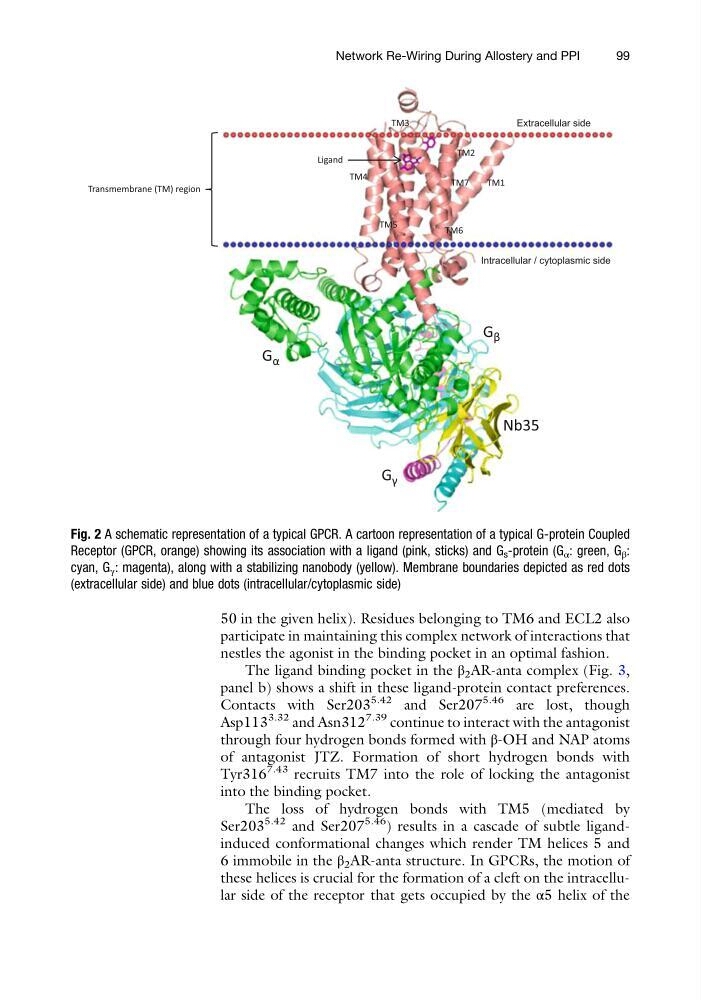
Fig. 2 A schematic representation of a typical GPCR. A cartoon representation of a typical G-protein Coupled
Receptor (GPCR, orange) showing its association with a ligand (pink, sticks) and G s -protein (G α : green, G β :
cyan, G γ : magenta), along with a stabilizing nanobody (yellow). Membrane boundaries depicted as red dots
(extracellular side) and blue dots (intracellular/cytoplasmic side)
Network Re-Wiring During Allostery and PPI
99
participate in maintaining this complex network of interactions that
nestles the agonist in the binding pocket in an optimal fashion.
The ligand binding pocket in the β 2 AR-anta complex (Fig. 3,
panel b) shows a shift in these ligand-protein contact preferences.
Contacts with Ser203
5.42 and Ser207
5.46 are lost, though
Asp113
3.32 and Asn312
7.39 continue to interact with the antagonist
through four hydrogen bonds formed with β-OH and NAP atoms
of antagonist JTZ. Formation of short hydrogen bonds with
Tyr316
7.43 recruits TM7 into the role of locking the antagonist
into the binding pocket.
The loss of hydrogen bonds with TM5 (mediated by
Ser203
5.42 and Ser207
5.46
) results in a cascade of subtle ligandinduced conformational changes which render TM helices 5 and
6 immobile in the β 2 AR-anta structure. In GPCRs, the motion of
these helices is crucial for the formation of a cleft on the intracellular side of the receptor that gets occupied by the α5 helix of the
Extracellular side
Intracellular / cytoplasmic side
G α
G β
G γ
Nb35
Transmembrane (TM) region
TM1
TM2
TM3
TM4
TM5
TM6
TM7
Ligand
Fig. 2 A schematic representation of a typical GPCR. A cartoon representation of a typical G-protein Coupled
Receptor (GPCR, orange) showing its association with a ligand (pink, sticks) and G s -protein (G α : green, G β :
cyan, G γ : magenta), along with a stabilizing nanobody (yellow). Membrane boundaries depicted as red dots
(extracellular side) and blue dots (intracellular/cytoplasmic side)
Network Re-Wiring During Allostery and PPI
99