

2. Extract genomic DNA directly in the wells using 25 μL of
commercially available DNA lysis buffer.
3. Proceed to PCR amplification from genomic DNA of each
clone with the primer pairs XPB-FW/XPB-RV or XPB-FW/
GFP-RV following the conditions used for PCR analysis of the
pool of transfected cells. Run 2 μL of the PCR on 1% agarose
TBE 1Â gel at 100 V for 1 h. Visualize the PCR products using
a UV transilluminator (see Note 5).
4. Sequence the PCR fragments of clones to ensure correct tag
sequence insertion.
3.5.2 Western Blot
Analysis
1. Seed five 150-mm Petri dishes containing 25 mL of growth
medium and incubate at 37
C, 5% CO 2 until a confluency of
80% is reached.
2. Wash the plate with PBS containing 30% w/v glycerol, detach
the cells with a scraper, and after centrifugation at 1000 Â g for
10 min at 4
C, snap freeze the pellet in liquid nitrogen and
store at À80
C. We usually prepare batches of 15 Â 10
6 and
25 Â 10
6 cells.
3. Resuspend 15 Â 10
6 cells in 150 μL of RIPA buffer and
incubate for 10 min by pipetting up and down.
A
B
XPB-GFP
XPB
250
130
70
55
35
25
1
2
U 2 O S : X P B - G F P
U 2 O S
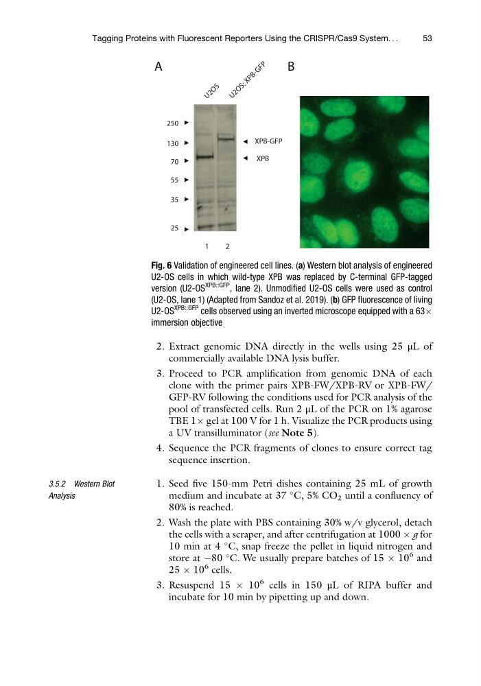
Fig. 6 Validation of engineered cell lines. (a) Western blot analysis of engineered
U2-OS cells in which wild-type XPB was replaced by C-terminal GFP-tagged
version (U2-OS
XPB::GFP
, lane 2). Unmodified U2-OS cells were used as control
(U2-OS, lane 1) (Adapted from Sandoz et al. 2019). (b) GFP fluorescence of living
U2-OS
XPB::GFP
cells observed using an inverted microscope equipped with a 63Â
immersion objective
Tagging Proteins with Fluorescent Reporters Using the CRISPR/Cas9 System. . .
53
commercially available DNA lysis buffer.
3. Proceed to PCR amplification from genomic DNA of each
clone with the primer pairs XPB-FW/XPB-RV or XPB-FW/
GFP-RV following the conditions used for PCR analysis of the
pool of transfected cells. Run 2 μL of the PCR on 1% agarose
TBE 1Â gel at 100 V for 1 h. Visualize the PCR products using
a UV transilluminator (see Note 5).
4. Sequence the PCR fragments of clones to ensure correct tag
sequence insertion.
3.5.2 Western Blot
Analysis
1. Seed five 150-mm Petri dishes containing 25 mL of growth
medium and incubate at 37
C, 5% CO 2 until a confluency of
80% is reached.
2. Wash the plate with PBS containing 30% w/v glycerol, detach
the cells with a scraper, and after centrifugation at 1000 Â g for
10 min at 4
C, snap freeze the pellet in liquid nitrogen and
store at À80
C. We usually prepare batches of 15 Â 10
6 and
25 Â 10
6 cells.
3. Resuspend 15 Â 10
6 cells in 150 μL of RIPA buffer and
incubate for 10 min by pipetting up and down.
A
B
XPB-GFP
XPB
250
130
70
55
35
25
1
2
U 2 O S : X P B - G F P
U 2 O S
Fig. 6 Validation of engineered cell lines. (a) Western blot analysis of engineered
U2-OS cells in which wild-type XPB was replaced by C-terminal GFP-tagged
version (U2-OS
XPB::GFP
, lane 2). Unmodified U2-OS cells were used as control
(U2-OS, lane 1) (Adapted from Sandoz et al. 2019). (b) GFP fluorescence of living
U2-OS
XPB::GFP
cells observed using an inverted microscope equipped with a 63Â
immersion objective
Tagging Proteins with Fluorescent Reporters Using the CRISPR/Cas9 System. . .
53