

800 rpm for 15 min on a thermomixer, then immediately
remove the beads on the magnetic rack and transfer the eluted
material in a fresh 1.5-mL tube.
13. Eluted samples can be stored at À20
C until further
processing.
3.5 SDS-PAGE
and Sample
Preparation for MS
Analysis
1. Prior to sample preparation for MS analysis, assess the success
of the split-BioID experiment by Western blot. To do so,
prepare input and elution blot samples as follows: equal protein
amounts of input samples mixed with 5Â SDS loading buffer in
a total volume of 28 μL (load 25 μL per lane), 5 μL of each
elution sample mixed with 1.25 μL of 5Â SDS loading buffer
(load 6 μL per lane). Western blot analysis is performed as
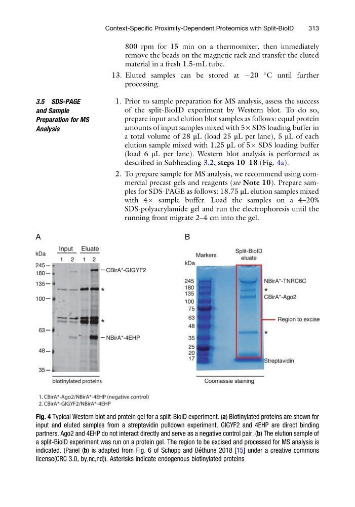
described in Subheading 3.2, steps 10–18 (Fig. 4a).
2. To prepare sample for MS analysis, we recommend using commercial precast gels and reagents (see Note 10). Prepare samples for SDS-PAGE as follows: 18.75 μL elution samples mixed
with 4Â sample buffer. Load the samples on a 4–20%
SDS-polyacrylamide gel and run the electrophoresis until the
running front migrate 2–4 cm into the gel.
Fig. 4 Typical Western blot and protein gel for a split-BioID experiment. (a) Biotinylated proteins are shown for
input and eluted samples from a streptavidin pulldown experiment. GIGYF2 and 4EHP are direct binding
partners. Ago2 and 4EHP do not interact directly and serve as a negative control pair. (b) The elution sample of
a split-BioID experiment was run on a protein gel. The region to be excised and processed for MS analysis is
indicated. (Panel (b) is adapted from Fig. 6 of Schopp and Be ´ thune 2018 [15] under a creative commons
license(CRC 3.0, by,nc,nd)). Asterisks indicate endogenous biotinylated proteins
Context-Specific Proximity-Dependent Proteomics with Split-BioID
313
remove the beads on the magnetic rack and transfer the eluted
material in a fresh 1.5-mL tube.
13. Eluted samples can be stored at À20
C until further
processing.
3.5 SDS-PAGE
and Sample
Preparation for MS
Analysis
1. Prior to sample preparation for MS analysis, assess the success
of the split-BioID experiment by Western blot. To do so,
prepare input and elution blot samples as follows: equal protein
amounts of input samples mixed with 5Â SDS loading buffer in
a total volume of 28 μL (load 25 μL per lane), 5 μL of each
elution sample mixed with 1.25 μL of 5Â SDS loading buffer
(load 6 μL per lane). Western blot analysis is performed as
described in Subheading 3.2, steps 10–18 (Fig. 4a).
2. To prepare sample for MS analysis, we recommend using commercial precast gels and reagents (see Note 10). Prepare samples for SDS-PAGE as follows: 18.75 μL elution samples mixed
with 4Â sample buffer. Load the samples on a 4–20%
SDS-polyacrylamide gel and run the electrophoresis until the
running front migrate 2–4 cm into the gel.
Fig. 4 Typical Western blot and protein gel for a split-BioID experiment. (a) Biotinylated proteins are shown for
input and eluted samples from a streptavidin pulldown experiment. GIGYF2 and 4EHP are direct binding
partners. Ago2 and 4EHP do not interact directly and serve as a negative control pair. (b) The elution sample of
a split-BioID experiment was run on a protein gel. The region to be excised and processed for MS analysis is
indicated. (Panel (b) is adapted from Fig. 6 of Schopp and Be ´ thune 2018 [15] under a creative commons
license(CRC 3.0, by,nc,nd)). Asterisks indicate endogenous biotinylated proteins
Context-Specific Proximity-Dependent Proteomics with Split-BioID
313