

pipetting up and down. The PCR tubes were placed into a Veriti™
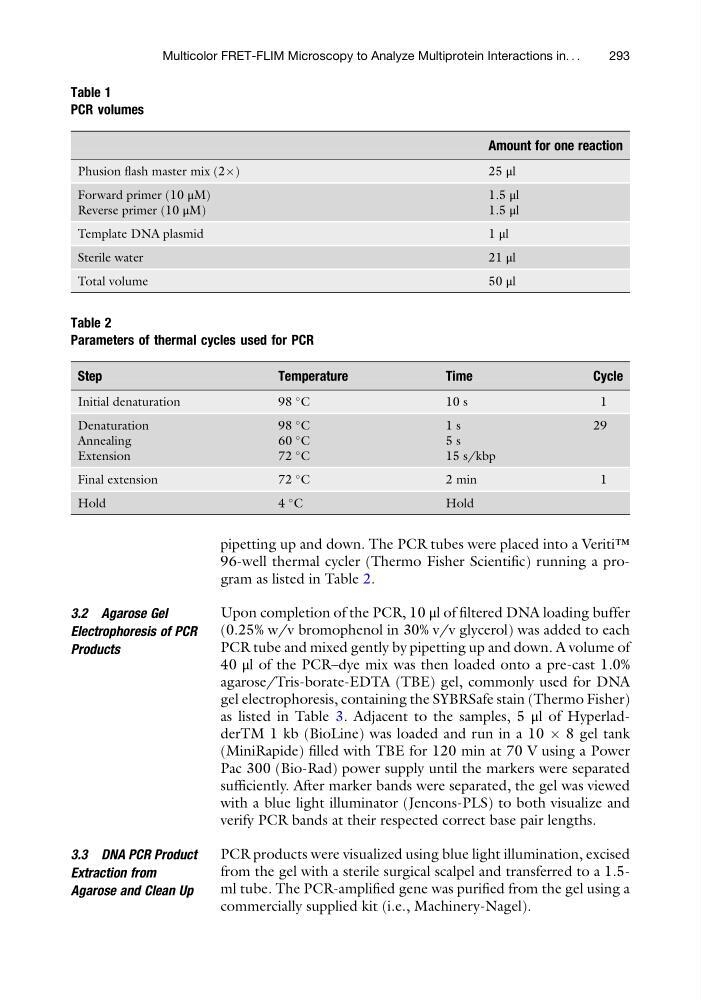
96-well thermal cycler (Thermo Fisher Scientific) running a program as listed in Table 2.
3.2 Agarose Gel
Electrophoresis of PCR
Products
Upon completion of the PCR, 10 μl of filtered DNA loading buffer
(0.25% w/v bromophenol in 30% v/v glycerol) was added to each
PCR tube and mixed gently by pipetting up and down. A volume of
40 μl of the PCR–dye mix was then loaded onto a pre-cast 1.0%
agarose/Tris-borate-EDTA (TBE) gel, commonly used for DNA
gel electrophoresis, containing the SYBRSafe stain (Thermo Fisher)
as listed in Table 3. Adjacent to the samples, 5 μl of HyperladderTM 1 kb (BioLine) was loaded and run in a 10 Â 8 gel tank
(MiniRapide) filled with TBE for 120 min at 70 V using a Power
Pac 300 (Bio-Rad) power supply until the markers were separated
sufficiently. After marker bands were separated, the gel was viewed
with a blue light illuminator (Jencons-PLS) to both visualize and
verify PCR bands at their respected correct base pair lengths.
3.3 DNA PCR Product
Extraction from
Agarose and Clean Up
PCR products were visualized using blue light illumination, excised
from the gel with a sterile surgical scalpel and transferred to a 1.5ml tube. The PCR-amplified gene was purified from the gel using a
commercially supplied kit (i.e., Machinery-Nagel).
Table 1
PCR volumes
Amount for one reaction
Phusion flash master mix (2Â)
2 5 μl
Forward primer (10 μM)
Reverse primer (10 μM)
1.5 μl
1.5 μl
Template DNA plasmid
1 μl
Sterile water
21 μl
Total volume
50 μl
Table 2
Parameters of thermal cycles used for PCR
Step
Temperature
Time
Cycle
Initial denaturation
98
C
1 0 s
1
Denaturation
Annealing
Extension
98
C
60
C
72
C
1 s
5 s
15 s/kbp
29
Final extension
72
C
2 min
1
Hold
4
C
Hold
Multicolor FRET-FLIM Microscopy to Analyze Multiprotein Interactions in. . .
293
96-well thermal cycler (Thermo Fisher Scientific) running a program as listed in Table 2.
3.2 Agarose Gel
Electrophoresis of PCR
Products
Upon completion of the PCR, 10 μl of filtered DNA loading buffer
(0.25% w/v bromophenol in 30% v/v glycerol) was added to each
PCR tube and mixed gently by pipetting up and down. A volume of
40 μl of the PCR–dye mix was then loaded onto a pre-cast 1.0%
agarose/Tris-borate-EDTA (TBE) gel, commonly used for DNA
gel electrophoresis, containing the SYBRSafe stain (Thermo Fisher)
as listed in Table 3. Adjacent to the samples, 5 μl of HyperladderTM 1 kb (BioLine) was loaded and run in a 10 Â 8 gel tank
(MiniRapide) filled with TBE for 120 min at 70 V using a Power
Pac 300 (Bio-Rad) power supply until the markers were separated
sufficiently. After marker bands were separated, the gel was viewed
with a blue light illuminator (Jencons-PLS) to both visualize and
verify PCR bands at their respected correct base pair lengths.
3.3 DNA PCR Product
Extraction from
Agarose and Clean Up
PCR products were visualized using blue light illumination, excised
from the gel with a sterile surgical scalpel and transferred to a 1.5ml tube. The PCR-amplified gene was purified from the gel using a
commercially supplied kit (i.e., Machinery-Nagel).
Table 1
PCR volumes
Amount for one reaction
Phusion flash master mix (2Â)
2 5 μl
Forward primer (10 μM)
Reverse primer (10 μM)
1.5 μl
1.5 μl
Template DNA plasmid
1 μl
Sterile water
21 μl
Total volume
50 μl
Table 2
Parameters of thermal cycles used for PCR
Step
Temperature
Time
Cycle
Initial denaturation
98
C
1 0 s
1
Denaturation
Annealing
Extension
98
C
60
C
72
C
1 s
5 s
15 s/kbp
29
Final extension
72
C
2 min
1
Hold
4
C
Hold
Multicolor FRET-FLIM Microscopy to Analyze Multiprotein Interactions in. . .
293