

During FRET, the rate of decay is reduced by a quenching
process that depletes the excited state of the donor fluorophore,
i.e., the donor fluorescence lifetime is shortened. Thus, by measuring changes in the excited-state lifetime of the donor at each pixel
making up an image, steady-state FRET can be more accurately
determined. This is described as fluorescence lifetime imaging
microscopy or FRET-FLIM [18–22]. Generally, the two proteins
under investigation are tagged with green fluorescent protein
(GFP) and its variants. In our work, we have used a combination
of blue FP (mTurquoise), yellow FP, and red FP (mCherry) as
donors and acceptors to investigate some of the multiple complex
mTORC1 proteins. We have also applied the FLIM imaging
method to investigate DNA structure as well as protein–protein
interactions in several systems including mammalians, viruses, and
plants [23–27], thus demonstrating wide applicability of FRETFLIM. We describe here a multi-color FRET-FLIM microscopy of
two-channel pulsed interleave excitation (PIE) to analyze multiprotein interactions in live cells.
Measuring the change in donor(s) lifetime in a FLIM experiment provides an elegant way to answer biological questions as well
as protein signaling that may involve multiple proteins at the same
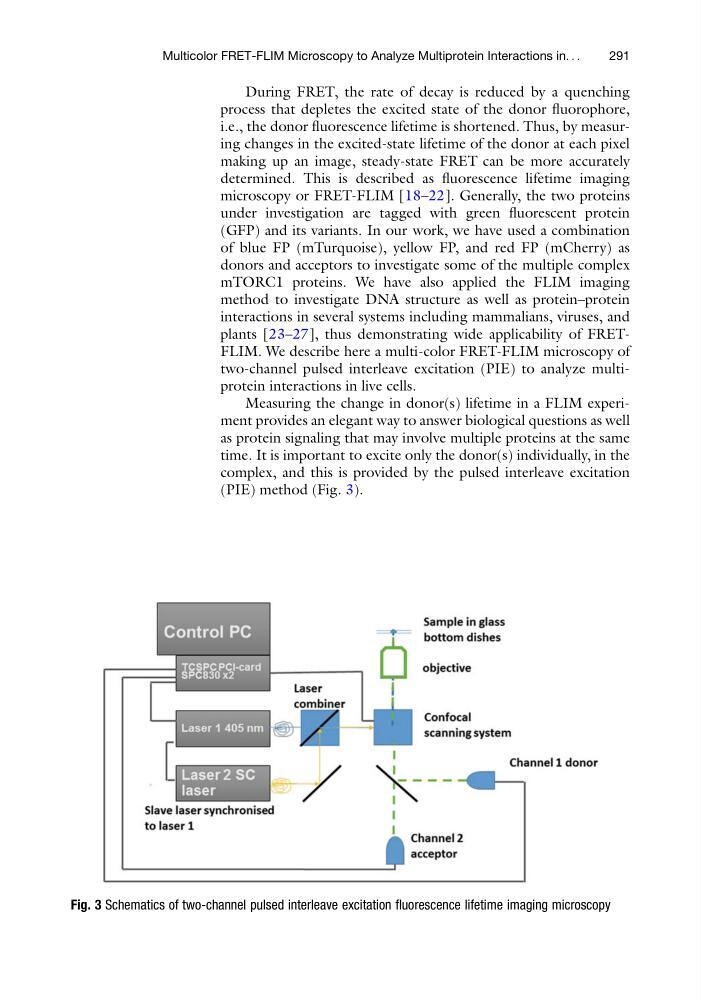
time. It is important to excite only the donor(s) individually, in the
complex, and this is provided by the pulsed interleave excitation
(PIE) method (Fig. 3).
Fig. 3 Schematics of two-channel pulsed interleave excitation fluorescence lifetime imaging microscopy
Multicolor FRET-FLIM Microscopy to Analyze Multiprotein Interactions in. . .
291
process that depletes the excited state of the donor fluorophore,
i.e., the donor fluorescence lifetime is shortened. Thus, by measuring changes in the excited-state lifetime of the donor at each pixel
making up an image, steady-state FRET can be more accurately
determined. This is described as fluorescence lifetime imaging
microscopy or FRET-FLIM [18–22]. Generally, the two proteins
under investigation are tagged with green fluorescent protein
(GFP) and its variants. In our work, we have used a combination
of blue FP (mTurquoise), yellow FP, and red FP (mCherry) as
donors and acceptors to investigate some of the multiple complex
mTORC1 proteins. We have also applied the FLIM imaging
method to investigate DNA structure as well as protein–protein
interactions in several systems including mammalians, viruses, and
plants [23–27], thus demonstrating wide applicability of FRETFLIM. We describe here a multi-color FRET-FLIM microscopy of
two-channel pulsed interleave excitation (PIE) to analyze multiprotein interactions in live cells.
Measuring the change in donor(s) lifetime in a FLIM experiment provides an elegant way to answer biological questions as well
as protein signaling that may involve multiple proteins at the same
time. It is important to excite only the donor(s) individually, in the
complex, and this is provided by the pulsed interleave excitation
(PIE) method (Fig. 3).
Fig. 3 Schematics of two-channel pulsed interleave excitation fluorescence lifetime imaging microscopy
Multicolor FRET-FLIM Microscopy to Analyze Multiprotein Interactions in. . .
291