

chaperones to facilitate crystallization, and as probes for cellular
imaging [8–30].
Phage display remains the most widely used approach for isolating protein binding reagents, despite the growing number of
alternative methods such as ribosome display [31], mRNA display
[32], and cis display [33]. Phage display was first described by
George P. Smith in 1985 [34] and has since been adapted to display
large libraries of antibody fragments, peptides, and artificial binding
proteins. Large libraries of Affimer reagents have been produced
that display Affimer proteins on a truncated pIII coat protein on
filamentous (Ff) phage [8]. Affimer reagents that specifically bind a
wide range of target proteins have been successfully isolated from
these libraries by phage display.
Selection of Affimer reagents by phage display is usually conducted over three panning rounds. Biotinylated target is immobilized on a streptavidin- or NeutrAvidin-coated surface and
incubated with a population of Affimer-displaying phage. After
washing away unbound phage, bound phage is eluted and propagated for subsequent rounds of panning. A typical method of
Affimer selection by phage display is depicted in Fig. 2.
The described method has been used to isolate isoform-specific
Affimer reagents [24, 25, 35]. The population of phage is first
incubated with the homologous protein to remove binders to the
isoform, prior to selection against the target protein (as depicted in
Fig. 2). We have also used this method of negative selection to
isolate specific binders of targets expressed as fusion proteins
InteracƟon
Loop 1
InteracƟon
Loop 2
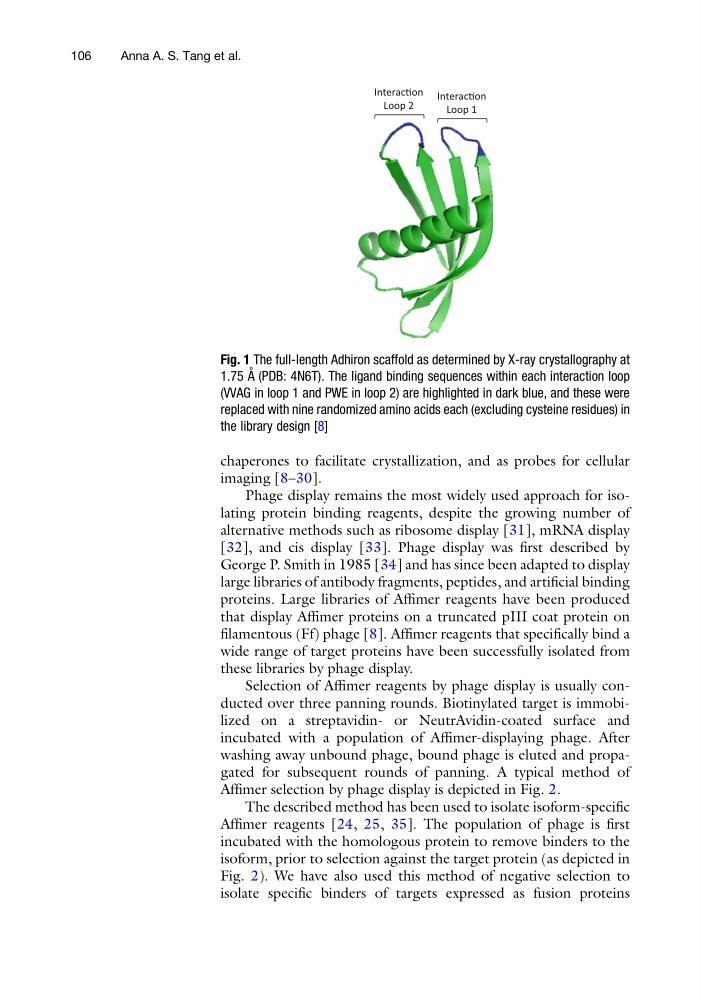
Fig. 1 The full-length Adhiron scaffold as determined by X-ray crystallography at
1.75 A ˚ (PDB: 4N6T). The ligand binding sequences within each interaction loop
(VVAG in loop 1 and PWE in loop 2) are highlighted in dark blue, and these were
replaced with nine randomized amino acids each (excluding cysteine residues) in
the library design [8]
106
Anna A. S. Tang et al.
imaging [8–30].
Phage display remains the most widely used approach for isolating protein binding reagents, despite the growing number of
alternative methods such as ribosome display [31], mRNA display
[32], and cis display [33]. Phage display was first described by
George P. Smith in 1985 [34] and has since been adapted to display
large libraries of antibody fragments, peptides, and artificial binding
proteins. Large libraries of Affimer reagents have been produced
that display Affimer proteins on a truncated pIII coat protein on
filamentous (Ff) phage [8]. Affimer reagents that specifically bind a
wide range of target proteins have been successfully isolated from
these libraries by phage display.
Selection of Affimer reagents by phage display is usually conducted over three panning rounds. Biotinylated target is immobilized on a streptavidin- or NeutrAvidin-coated surface and
incubated with a population of Affimer-displaying phage. After
washing away unbound phage, bound phage is eluted and propagated for subsequent rounds of panning. A typical method of
Affimer selection by phage display is depicted in Fig. 2.
The described method has been used to isolate isoform-specific
Affimer reagents [24, 25, 35]. The population of phage is first
incubated with the homologous protein to remove binders to the
isoform, prior to selection against the target protein (as depicted in
Fig. 2). We have also used this method of negative selection to
isolate specific binders of targets expressed as fusion proteins
InteracƟon
Loop 1
InteracƟon
Loop 2
Fig. 1 The full-length Adhiron scaffold as determined by X-ray crystallography at
1.75 A ˚ (PDB: 4N6T). The ligand binding sequences within each interaction loop
(VVAG in loop 1 and PWE in loop 2) are highlighted in dark blue, and these were
replaced with nine randomized amino acids each (excluding cysteine residues) in
the library design [8]
106
Anna A. S. Tang et al.