

60
J. Kumar V. and K. A. Reddy
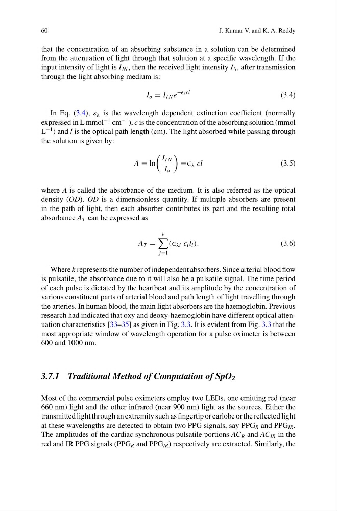
that the concentration of an absorbing substance in a solution can be determined
from the attenuation of light through that solution at a specific wavelength. If the
input intensity of light is I IN , then the received light intensity I 0 , after transmission
through the light absorbing medium is:
I o = I I N e
− λ cl
(3.4)
In Eq. (3.4), ε λ is the wavelength dependent extinction coefficient (normally
expressed in L mmol
−1 cm
−1 ), c is the concentration of the absorbing solution (mmol
L
−1 ) and l is the optical path length (cm). The light absorbed while passing through
the solution is given by:
A = ln
I I N
I o
=∈ λ cl
(3.5)
where A is called the absorbance of the medium. It is also referred as the optical
density (OD). OD is a dimensionless quantity. If multiple absorbers are present
in the path of light, then each absorber contributes its part and the resulting total
absorbance A T can be expressed as
A T =
k
j=1
(∈ λi c i l i ).
(3.6)
Where k represents the number of independent absorbers. Since arterial blood flow
is pulsatile, the absorbance due to it will also be a pulsatile signal. The time period
of each pulse is dictated by the heartbeat and its amplitude by the concentration of
various constituent parts of arterial blood and path length of light travelling through
the arteries. In human blood, the main light absorbers are the haemoglobin. Previous
research had indicated that oxy and deoxy-haemoglobin have different optical attenuation characteristics [33–35] as given in Fig. 3.3. It is evident from Fig. 3.3 that the
most appropriate window of wavelength operation for a pulse oximeter is between
600 and 1000 nm.
3.7.1 Traditional Method of Computation of SpO 2
Most of the commercial pulse oximeters employ two LEDs, one emitting red (near
660 nm) light and the other infrared (near 900 nm) light as the sources. Either the
transmitted light through an extremity such as fingertip or earlobe or the reflected light
at these wavelengths are detected to obtain two PPG signals, say PPG R and PPG IR .
The amplitudes of the cardiac synchronous pulsatile portions AC R and AC IR in the
red and IR PPG signals (PPG R and PPG IR ) respectively are extracted. Similarly, the
J. Kumar V. and K. A. Reddy
that the concentration of an absorbing substance in a solution can be determined
from the attenuation of light through that solution at a specific wavelength. If the
input intensity of light is I IN , then the received light intensity I 0 , after transmission
through the light absorbing medium is:
I o = I I N e
− λ cl
(3.4)
In Eq. (3.4), ε λ is the wavelength dependent extinction coefficient (normally
expressed in L mmol
−1 cm
−1 ), c is the concentration of the absorbing solution (mmol
L
−1 ) and l is the optical path length (cm). The light absorbed while passing through
the solution is given by:
A = ln
I I N
I o
=∈ λ cl
(3.5)
where A is called the absorbance of the medium. It is also referred as the optical
density (OD). OD is a dimensionless quantity. If multiple absorbers are present
in the path of light, then each absorber contributes its part and the resulting total
absorbance A T can be expressed as
A T =
k
j=1
(∈ λi c i l i ).
(3.6)
Where k represents the number of independent absorbers. Since arterial blood flow
is pulsatile, the absorbance due to it will also be a pulsatile signal. The time period
of each pulse is dictated by the heartbeat and its amplitude by the concentration of
various constituent parts of arterial blood and path length of light travelling through
the arteries. In human blood, the main light absorbers are the haemoglobin. Previous
research had indicated that oxy and deoxy-haemoglobin have different optical attenuation characteristics [33–35] as given in Fig. 3.3. It is evident from Fig. 3.3 that the
most appropriate window of wavelength operation for a pulse oximeter is between
600 and 1000 nm.
3.7.1 Traditional Method of Computation of SpO 2
Most of the commercial pulse oximeters employ two LEDs, one emitting red (near
660 nm) light and the other infrared (near 900 nm) light as the sources. Either the
transmitted light through an extremity such as fingertip or earlobe or the reflected light
at these wavelengths are detected to obtain two PPG signals, say PPG R and PPG IR .
The amplitudes of the cardiac synchronous pulsatile portions AC R and AC IR in the
red and IR PPG signals (PPG R and PPG IR ) respectively are extracted. Similarly, the