

2 Influence of Controlled Breathing (Pranayama) on Dermal Perfusion
39
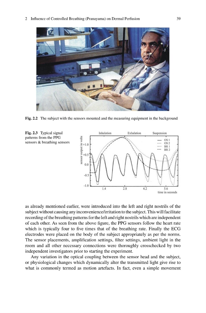
Fig. 2.2 The subject with the sensors mounted and the measuring equipment in the background
Fig. 2.3 Typical signal
patterns from the PPG
sensors & breathing sensors
as already mentioned earlier, were introduced into the left and right nostrils of the
subject without causing any inconvenience/irritation to the subject. This will facilitate
recording of the breathing patterns for the left and right nostrils which are independent
of each other. As seen from the above figure, the PPG sensors follow the heart rate
which is typically four to five times that of the breathing rate. Finally the ECG
electrodes were placed on the body of the subject appropriately as per the norms.
The sensor placements, amplification settings, filter settings, ambient light in the
room and all other necessary connections were thoroughly crosschecked by two
independent investigators prior to starting the experiment.
Any variation in the optical coupling between the sensor head and the subject,
or physiological changes which dynamically alter the transmitted light give rise to
what is commonly termed as motion artefacts. In fact, even a simple movement
39
Fig. 2.2 The subject with the sensors mounted and the measuring equipment in the background
Fig. 2.3 Typical signal
patterns from the PPG
sensors & breathing sensors
as already mentioned earlier, were introduced into the left and right nostrils of the
subject without causing any inconvenience/irritation to the subject. This will facilitate
recording of the breathing patterns for the left and right nostrils which are independent
of each other. As seen from the above figure, the PPG sensors follow the heart rate
which is typically four to five times that of the breathing rate. Finally the ECG
electrodes were placed on the body of the subject appropriately as per the norms.
The sensor placements, amplification settings, filter settings, ambient light in the
room and all other necessary connections were thoroughly crosschecked by two
independent investigators prior to starting the experiment.
Any variation in the optical coupling between the sensor head and the subject,
or physiological changes which dynamically alter the transmitted light give rise to
what is commonly termed as motion artefacts. In fact, even a simple movement