

1 Skin Perfusion Studies: Historical Notes and Modern Measuring …
13
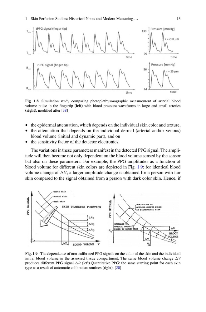
Fig. 1.8 Simulation study comparing photoplethysmographic measurement of arterial blood
volume pulse in the fingertip (left) with blood pressure waveforms in large and small arteries
(right), modified after [38]
• the epidermal attenuation, which depends on the individual skin color and texture,
• the attenuation that depends on the individual dermal (arterial and/or venous)
blood volume (initial and dynamic part), and on
• the sensitivity factor of the detector electronics.
The variations in these parameters manifest in the detected PPG signal. The amplitude will then become not only dependent on the blood volume sensed by the sensor
but also on these parameters. For example, the PPG amplitudes as a function of
blood volume for different skin colors are depicted in Fig. 1.9: for identical blood
volume change of V, a larger amplitude change is obtained for a person with fair
skin compared to the signal obtained from a person with dark color skin. Hence, if
Fig. 1.9 The dependence of non-calibrated PPG signals on the color of the skin and the individual
initial blood volume in the assessed tissue compartment. The same blood volume change V
produces different PPG signal R (left).Quantitative PPG: the same starting point for each skin
type as a result of automatic calibration routines (right), [20]
13
Fig. 1.8 Simulation study comparing photoplethysmographic measurement of arterial blood
volume pulse in the fingertip (left) with blood pressure waveforms in large and small arteries
(right), modified after [38]
• the epidermal attenuation, which depends on the individual skin color and texture,
• the attenuation that depends on the individual dermal (arterial and/or venous)
blood volume (initial and dynamic part), and on
• the sensitivity factor of the detector electronics.
The variations in these parameters manifest in the detected PPG signal. The amplitude will then become not only dependent on the blood volume sensed by the sensor
but also on these parameters. For example, the PPG amplitudes as a function of
blood volume for different skin colors are depicted in Fig. 1.9: for identical blood
volume change of V, a larger amplitude change is obtained for a person with fair
skin compared to the signal obtained from a person with dark color skin. Hence, if
Fig. 1.9 The dependence of non-calibrated PPG signals on the color of the skin and the individual
initial blood volume in the assessed tissue compartment. The same blood volume change V
produces different PPG signal R (left).Quantitative PPG: the same starting point for each skin
type as a result of automatic calibration routines (right), [20]