

13 Concluding Remarks and New Horizons in Skin Perfusion Studies
229
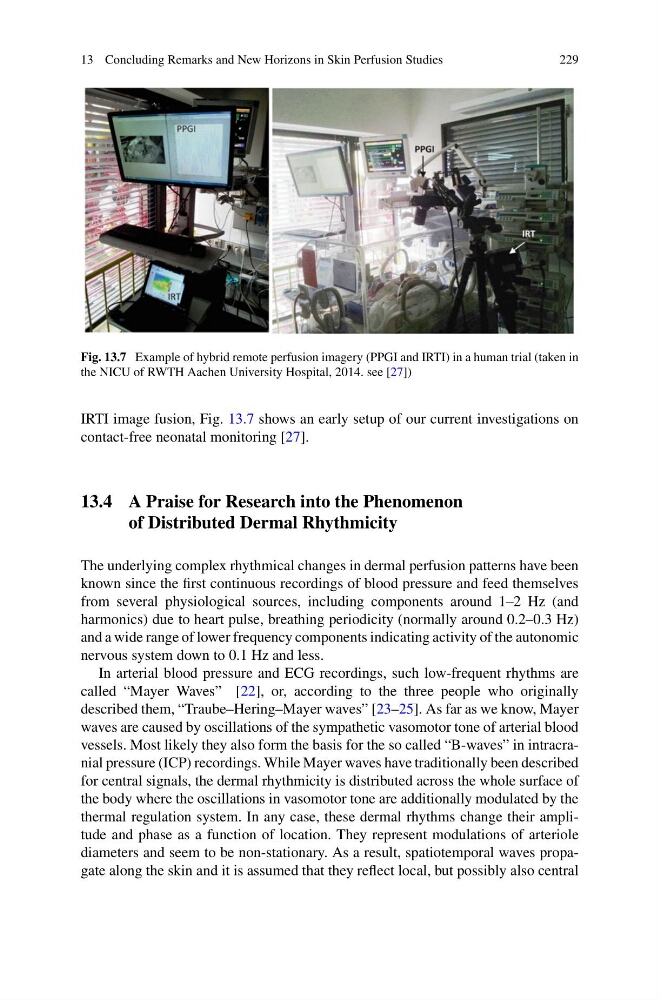
Fig. 13.7 Example of hybrid remote perfusion imagery (PPGI and IRTI) in a human trial (taken in
the NICU of RWTH Aachen University Hospital, 2014. see [27])
IRTI image fusion, Fig. 13.7 shows an early setup of our current investigations on
contact-free neonatal monitoring [27].
13.4 A Praise for Research into the Phenomenon
of Distributed Dermal Rhythmicity
The underlying complex rhythmical changes in dermal perfusion patterns have been
known since the first continuous recordings of blood pressure and feed themselves
from several physiological sources, including components around 1–2 Hz (and
harmonics) due to heart pulse, breathing periodicity (normally around 0.2–0.3 Hz)
and a wide range of lower frequency components indicating activity of the autonomic
nervous system down to 0.1 Hz and less.
In arterial blood pressure and ECG recordings, such low-frequent rhythms are
called “Mayer Waves” [22], or, according to the three people who originally
described them, “Traube–Hering–Mayer waves” [23–25]. As far as we know, Mayer
waves are caused by oscillations of the sympathetic vasomotor tone of arterial blood
vessels. Most likely they also form the basis for the so called “B-waves” in intracranial pressure (ICP) recordings. While Mayer waves have traditionally been described
for central signals, the dermal rhythmicity is distributed across the whole surface of
the body where the oscillations in vasomotor tone are additionally modulated by the
thermal regulation system. In any case, these dermal rhythms change their amplitude and phase as a function of location. They represent modulations of arteriole
diameters and seem to be non-stationary. As a result, spatiotemporal waves propagate along the skin and it is assumed that they reflect local, but possibly also central
229
Fig. 13.7 Example of hybrid remote perfusion imagery (PPGI and IRTI) in a human trial (taken in
the NICU of RWTH Aachen University Hospital, 2014. see [27])
IRTI image fusion, Fig. 13.7 shows an early setup of our current investigations on
contact-free neonatal monitoring [27].
13.4 A Praise for Research into the Phenomenon
of Distributed Dermal Rhythmicity
The underlying complex rhythmical changes in dermal perfusion patterns have been
known since the first continuous recordings of blood pressure and feed themselves
from several physiological sources, including components around 1–2 Hz (and
harmonics) due to heart pulse, breathing periodicity (normally around 0.2–0.3 Hz)
and a wide range of lower frequency components indicating activity of the autonomic
nervous system down to 0.1 Hz and less.
In arterial blood pressure and ECG recordings, such low-frequent rhythms are
called “Mayer Waves” [22], or, according to the three people who originally
described them, “Traube–Hering–Mayer waves” [23–25]. As far as we know, Mayer
waves are caused by oscillations of the sympathetic vasomotor tone of arterial blood
vessels. Most likely they also form the basis for the so called “B-waves” in intracranial pressure (ICP) recordings. While Mayer waves have traditionally been described
for central signals, the dermal rhythmicity is distributed across the whole surface of
the body where the oscillations in vasomotor tone are additionally modulated by the
thermal regulation system. In any case, these dermal rhythms change their amplitude and phase as a function of location. They represent modulations of arteriole
diameters and seem to be non-stationary. As a result, spatiotemporal waves propagate along the skin and it is assumed that they reflect local, but possibly also central