

190
N. Blanik
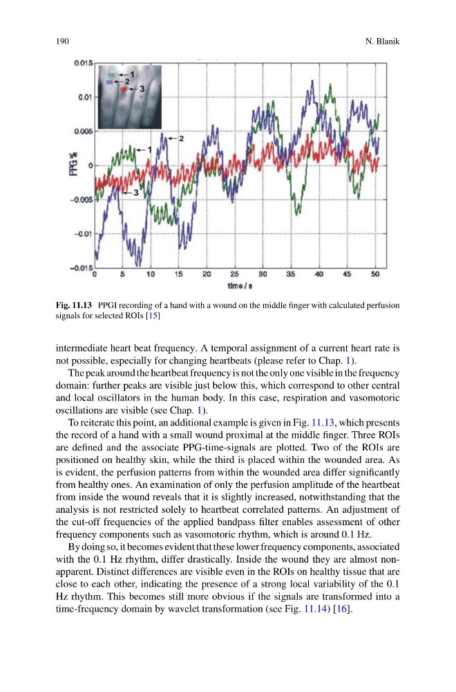
Fig. 11.13 PPGI recording of a hand with a wound on the middle finger with calculated perfusion
signals for selected ROIs [15]
intermediate heart beat frequency. A temporal assignment of a current heart rate is
not possible, especially for changing heartbeats (please refer to Chap. 1).
The peak around the heartbeat frequency is not the only one visible in the frequency
domain: further peaks are visible just below this, which correspond to other central
and local oscillators in the human body. In this case, respiration and vasomotoric
oscillations are visible (see Chap. 1).
To reiterate this point, an additional example is given in Fig. 11.13, which presents
the record of a hand with a small wound proximal at the middle finger. Three ROIs
are defined and the associate PPG-time-signals are plotted. Two of the ROIs are
positioned on healthy skin, while the third is placed within the wounded area. As
is evident, the perfusion patterns from within the wounded area differ significantly
from healthy ones. An examination of only the perfusion amplitude of the heartbeat
from inside the wound reveals that it is slightly increased, notwithstanding that the
analysis is not restricted solely to heartbeat correlated patterns. An adjustment of
the cut-off frequencies of the applied bandpass filter enables assessment of other
frequency components such as vasomotoric rhythm, which is around 0.1 Hz.
By doing so, it becomes evident that these lower frequency components, associated
with the 0.1 Hz rhythm, differ drastically. Inside the wound they are almost nonapparent. Distinct differences are visible even in the ROIs on healthy tissue that are
close to each other, indicating the presence of a strong local variability of the 0.1
Hz rhythm. This becomes still more obvious if the signals are transformed into a
time-frequency domain by wavelet transformation (see Fig. 11.14) [16].
N. Blanik
Fig. 11.13 PPGI recording of a hand with a wound on the middle finger with calculated perfusion
signals for selected ROIs [15]
intermediate heart beat frequency. A temporal assignment of a current heart rate is
not possible, especially for changing heartbeats (please refer to Chap. 1).
The peak around the heartbeat frequency is not the only one visible in the frequency
domain: further peaks are visible just below this, which correspond to other central
and local oscillators in the human body. In this case, respiration and vasomotoric
oscillations are visible (see Chap. 1).
To reiterate this point, an additional example is given in Fig. 11.13, which presents
the record of a hand with a small wound proximal at the middle finger. Three ROIs
are defined and the associate PPG-time-signals are plotted. Two of the ROIs are
positioned on healthy skin, while the third is placed within the wounded area. As
is evident, the perfusion patterns from within the wounded area differ significantly
from healthy ones. An examination of only the perfusion amplitude of the heartbeat
from inside the wound reveals that it is slightly increased, notwithstanding that the
analysis is not restricted solely to heartbeat correlated patterns. An adjustment of
the cut-off frequencies of the applied bandpass filter enables assessment of other
frequency components such as vasomotoric rhythm, which is around 0.1 Hz.
By doing so, it becomes evident that these lower frequency components, associated
with the 0.1 Hz rhythm, differ drastically. Inside the wound they are almost nonapparent. Distinct differences are visible even in the ROIs on healthy tissue that are
close to each other, indicating the presence of a strong local variability of the 0.1
Hz rhythm. This becomes still more obvious if the signals are transformed into a
time-frequency domain by wavelet transformation (see Fig. 11.14) [16].