

11 Remote Space- and Time-Resolved Skin Perfusion Detection …
181
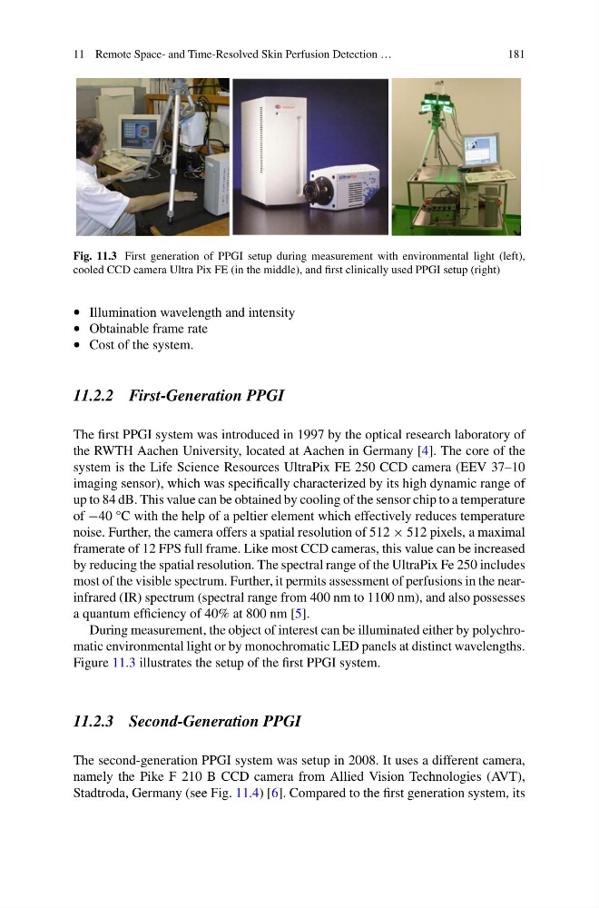
Fig. 11.3 First generation of PPGI setup during measurement with environmental light (left),
cooled CCD camera Ultra Pix FE (in the middle), and first clinically used PPGI setup (right)
• Illumination wavelength and intensity
• Obtainable frame rate
• Cost of the system.
11.2.2 First-Generation PPGI
The first PPGI system was introduced in 1997 by the optical research laboratory of
the RWTH Aachen University, located at Aachen in Germany [4]. The core of the
system is the Life Science Resources UltraPix FE 250 CCD camera (EEV 37–10
imaging sensor), which was specifically characterized by its high dynamic range of
up to 84 dB. This value can be obtained by cooling of the sensor chip to a temperature
of −40 °C with the help of a peltier element which effectively reduces temperature
noise. Further, the camera offers a spatial resolution of 512 × 512 pixels, a maximal
framerate of 12 FPS full frame. Like most CCD cameras, this value can be increased
by reducing the spatial resolution. The spectral range of the UltraPix Fe 250 includes
most of the visible spectrum. Further, it permits assessment of perfusions in the nearinfrared (IR) spectrum (spectral range from 400 nm to 1100 nm), and also possesses
a quantum efficiency of 40% at 800 nm [5].
During measurement, the object of interest can be illuminated either by polychromatic environmental light or by monochromatic LED panels at distinct wavelengths.
Figure 11.3 illustrates the setup of the first PPGI system.
11.2.3 Second-Generation PPGI
The second-generation PPGI system was setup in 2008. It uses a different camera,
namely the Pike F 210 B CCD camera from Allied Vision Technologies (AVT),
Stadtroda, Germany (see Fig. 11.4) [6]. Compared to the first generation system, its
181
Fig. 11.3 First generation of PPGI setup during measurement with environmental light (left),
cooled CCD camera Ultra Pix FE (in the middle), and first clinically used PPGI setup (right)
• Illumination wavelength and intensity
• Obtainable frame rate
• Cost of the system.
11.2.2 First-Generation PPGI
The first PPGI system was introduced in 1997 by the optical research laboratory of
the RWTH Aachen University, located at Aachen in Germany [4]. The core of the
system is the Life Science Resources UltraPix FE 250 CCD camera (EEV 37–10
imaging sensor), which was specifically characterized by its high dynamic range of
up to 84 dB. This value can be obtained by cooling of the sensor chip to a temperature
of −40 °C with the help of a peltier element which effectively reduces temperature
noise. Further, the camera offers a spatial resolution of 512 × 512 pixels, a maximal
framerate of 12 FPS full frame. Like most CCD cameras, this value can be increased
by reducing the spatial resolution. The spectral range of the UltraPix Fe 250 includes
most of the visible spectrum. Further, it permits assessment of perfusions in the nearinfrared (IR) spectrum (spectral range from 400 nm to 1100 nm), and also possesses
a quantum efficiency of 40% at 800 nm [5].
During measurement, the object of interest can be illuminated either by polychromatic environmental light or by monochromatic LED panels at distinct wavelengths.
Figure 11.3 illustrates the setup of the first PPGI system.
11.2.3 Second-Generation PPGI
The second-generation PPGI system was setup in 2008. It uses a different camera,
namely the Pike F 210 B CCD camera from Allied Vision Technologies (AVT),
Stadtroda, Germany (see Fig. 11.4) [6]. Compared to the first generation system, its