

apical dome, are depending on calcium is strongly supported by fluorescence
imaging demonstrating a drastic shift of the steep tip-high calcium gradient and
putative calcium channels towards the upper flank shortly before the relocalization
of the Spitzenkörper and the initiation of the graviresponse in protonemata (Figs. 4.8
and 4.9). Such a shift of the apical calcium gradient has never been observed in
rhizoids (Braun and Richter 1999).
Considering this wealth of data, it is tempting to speculate that the asymmetric
influx of calcium mediates the repositioning of the Spitzenkörper and the growth
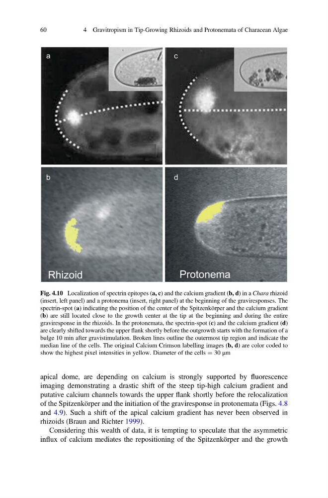
Fig. 4.10 Localization of spectrin epitopes (a, c) and the calcium gradient (b, d) in a Chara rhizoid
(insert, left panel) and a protonema (insert, right panel) at the beginning of the graviresponses. The
spectrin-spot (a) indicating the position of the center of the Spitzenkörper and the calcium gradient
(b) are still located close to the growth center at the tip at the beginning and during the entire
graviresponse in the rhizoids. In the protonemata, the spectrin-spot (c) and the calcium gradient (d)
are clearly shifted towards the upper flank shortly before the outgrowth starts with the formation of a
bulge 10 min after gravistimulation. Broken lines outline the outermost tip region and indicate the
median line of the cells. The original Calcium Crimson labelling images (b, d) are color coded to
show the highest pixel intensities in yellow. Diameter of the cells ¼ 30 μm
60
4 Gravitropism in Tip-Growing Rhizoids and Protonemata of Characean Algae
imaging demonstrating a drastic shift of the steep tip-high calcium gradient and
putative calcium channels towards the upper flank shortly before the relocalization
of the Spitzenkörper and the initiation of the graviresponse in protonemata (Figs. 4.8
and 4.9). Such a shift of the apical calcium gradient has never been observed in
rhizoids (Braun and Richter 1999).
Considering this wealth of data, it is tempting to speculate that the asymmetric
influx of calcium mediates the repositioning of the Spitzenkörper and the growth
Fig. 4.10 Localization of spectrin epitopes (a, c) and the calcium gradient (b, d) in a Chara rhizoid
(insert, left panel) and a protonema (insert, right panel) at the beginning of the graviresponses. The
spectrin-spot (a) indicating the position of the center of the Spitzenkörper and the calcium gradient
(b) are still located close to the growth center at the tip at the beginning and during the entire
graviresponse in the rhizoids. In the protonemata, the spectrin-spot (c) and the calcium gradient (d)
are clearly shifted towards the upper flank shortly before the outgrowth starts with the formation of a
bulge 10 min after gravistimulation. Broken lines outline the outermost tip region and indicate the
median line of the cells. The original Calcium Crimson labelling images (b, d) are color coded to
show the highest pixel intensities in yellow. Diameter of the cells ¼ 30 μm
60
4 Gravitropism in Tip-Growing Rhizoids and Protonemata of Characean Algae