

The negative graviresponse in protonema has been described as ´bending by
bulging´ (Hodick 1994) which refers to the bulge that appears on the upper cell flank
at the beginning of the graviresponse indicating a drastic relocation of the center of
maximal growth towards to upper flank (Fig. 4.3). And indeed, the Spitzenkörper
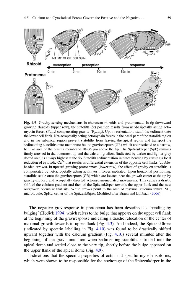
(indicated by spectrin labelling in Fig. 4.10) was found to be drastically shifted
upward together with the calcium gradient (Fig. 4.10) several minutes after the
beginning of the gravistimulation when sedimenting statoliths intruded into the
apical dome and settled close to the very tip, shortly before the bulge appeared on
the upper flank of the apical dome (Fig. 4.9).
Indications that the specific properties of actin and specific myosin isoforms,
which were shown to be responsible for the anchorage of the Spitzenkörper in the
Fig. 4.9 Gravity-sensing mechanisms in characean rhizoids and protonemata. In tip-downward
growing rhizoids (upper row), the statolith (St) position results from net-basipetally acting actomyosin forces (F actin ) compensating gravity (F gravity ). Upon reorientation, statoliths sediment onto
the lower cell flank. Net-acropetally acting actomyosin forces in the basal part of the statolith region
and in the subapical region prevent statoliths from leaving the apical region and transport the
sedimenting statoliths onto membrane-bound gravireceptors (GR) which are restricted to a narrow,
beltlike area of the plasma membrane 10–35 μm above the tip. The Spitzenkörper (Spk) remains
firmly arrested in the outermost tip and the calcium gradient (indicated by darker and lighter grey
dotted area) is always highest at the tip. Statolith sedimentation initiates bending by causing a local
reduction of cytosolic Ca
2+ that results in differential extension of the opposite cell flanks (doubleheaded arrows). In upward growing protonemata (lower row), the effect of gravity on statoliths is
compensated by net-acropetally acting actomyosin forces mediated. Upon horizontal positioning,
statoliths settle onto the gravireceptors (GR) which are located near the growth center at the tip by
gravity-induced and acropetally directed actomyosin-mediated movements. This causes a drastic
shift of the calcium gradient and then of the Spitzenkörper towards the upper flank and the new
outgrowth occurs at that site. White arrows point to the area of maximal calcium influx. MT,
microtubule; SpKc, center of the Spitzenkörper. Modified after Braun and Limbach (2006)
4.5 Calcium and Cytoskeletal Forces Govern the Positive and the Negative. . .
59
bulging´ (Hodick 1994) which refers to the bulge that appears on the upper cell flank
at the beginning of the graviresponse indicating a drastic relocation of the center of
maximal growth towards to upper flank (Fig. 4.3). And indeed, the Spitzenkörper
(indicated by spectrin labelling in Fig. 4.10) was found to be drastically shifted
upward together with the calcium gradient (Fig. 4.10) several minutes after the
beginning of the gravistimulation when sedimenting statoliths intruded into the
apical dome and settled close to the very tip, shortly before the bulge appeared on
the upper flank of the apical dome (Fig. 4.9).
Indications that the specific properties of actin and specific myosin isoforms,
which were shown to be responsible for the anchorage of the Spitzenkörper in the
Fig. 4.9 Gravity-sensing mechanisms in characean rhizoids and protonemata. In tip-downward
growing rhizoids (upper row), the statolith (St) position results from net-basipetally acting actomyosin forces (F actin ) compensating gravity (F gravity ). Upon reorientation, statoliths sediment onto
the lower cell flank. Net-acropetally acting actomyosin forces in the basal part of the statolith region
and in the subapical region prevent statoliths from leaving the apical region and transport the
sedimenting statoliths onto membrane-bound gravireceptors (GR) which are restricted to a narrow,
beltlike area of the plasma membrane 10–35 μm above the tip. The Spitzenkörper (Spk) remains
firmly arrested in the outermost tip and the calcium gradient (indicated by darker and lighter grey
dotted area) is always highest at the tip. Statolith sedimentation initiates bending by causing a local
reduction of cytosolic Ca
2+ that results in differential extension of the opposite cell flanks (doubleheaded arrows). In upward growing protonemata (lower row), the effect of gravity on statoliths is
compensated by net-acropetally acting actomyosin forces mediated. Upon horizontal positioning,
statoliths settle onto the gravireceptors (GR) which are located near the growth center at the tip by
gravity-induced and acropetally directed actomyosin-mediated movements. This causes a drastic
shift of the calcium gradient and then of the Spitzenkörper towards the upper flank and the new
outgrowth occurs at that site. White arrows point to the area of maximal calcium influx. MT,
microtubule; SpKc, center of the Spitzenkörper. Modified after Braun and Limbach (2006)
4.5 Calcium and Cytoskeletal Forces Govern the Positive and the Negative. . .
59