

by cytochalasin D or the calcium ionophore A23187, which both cause a disintegration
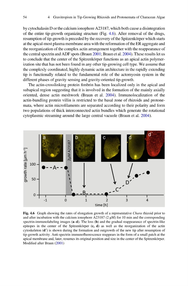
of the entire tip-growth organizing structure (Fig. 4.6). After removal of the drugs,
resumption of tip-growth is preceded by the recovery of the Spitzenkörper which starts
at the apical-most plasma membrane area with the reformation of the ER aggregate and
the reorganization of the complex actin arrangement together with the reappearance of
the central spectrin and ADF spots (Braun 2001; Braun et al. 2004). These results let us
to conclude that the center of the Spitzenkörper functions as an apical actin polymerization site that has not been found in any other tip-growing cell type. We assume that
the complexly coordinated, highly dynamic actin architecture in the rapidly extending
tip is functionally related to the fundamental role of the actomyosin system in the
different phases of gravity sensing and gravity-oriented tip-growth.
The actin-crosslinking protein fimbrin has been localized only in the apical and
subapical region suggesting that it is involved in the formation of the mainly axially
oriented, dense actin meshwork (Braun et al. 2004). Immunolocalization of the
actin-bundling protein villin is restricted to the basal zone of rhizoids and protonemata, where actin microfilaments are separated according to their polarity and form
two populations of thick interconnected actin bundles which generate the rotational
cytoplasmic streaming around the large central vacuole (Braun et al. 2004).
Fig. 4.6 Graph showing the rates of elongation growth of a representative Chara rhizoid prior to
and after incubation with the calcium ionophore A23187 (2 μM) for 10 min and the corresponding
spectrin-immunolabeling images (a–d). The loss (b) and the gradual reappearance of spectrin-like
epitopes in the center of the Spitzenkörper (c, d) as well as the reorganization of the actin
cytoskeleton (d´) is shown during the formation and outgrowth of the new tip after resumption of
tip-growth activity. Anti-spectrin immunofluorescence reappears in the form of a small patch at the
apical membrane and, later, resumes its original position and size in the center of the Spitzenkörper.
Modified after Braun (2001)
54
4 Gravitropism in Tip-Growing Rhizoids and Protonemata of Characean Algae
of the entire tip-growth organizing structure (Fig. 4.6). After removal of the drugs,
resumption of tip-growth is preceded by the recovery of the Spitzenkörper which starts
at the apical-most plasma membrane area with the reformation of the ER aggregate and
the reorganization of the complex actin arrangement together with the reappearance of
the central spectrin and ADF spots (Braun 2001; Braun et al. 2004). These results let us
to conclude that the center of the Spitzenkörper functions as an apical actin polymerization site that has not been found in any other tip-growing cell type. We assume that
the complexly coordinated, highly dynamic actin architecture in the rapidly extending
tip is functionally related to the fundamental role of the actomyosin system in the
different phases of gravity sensing and gravity-oriented tip-growth.
The actin-crosslinking protein fimbrin has been localized only in the apical and
subapical region suggesting that it is involved in the formation of the mainly axially
oriented, dense actin meshwork (Braun et al. 2004). Immunolocalization of the
actin-bundling protein villin is restricted to the basal zone of rhizoids and protonemata, where actin microfilaments are separated according to their polarity and form
two populations of thick interconnected actin bundles which generate the rotational
cytoplasmic streaming around the large central vacuole (Braun et al. 2004).
Fig. 4.6 Graph showing the rates of elongation growth of a representative Chara rhizoid prior to
and after incubation with the calcium ionophore A23187 (2 μM) for 10 min and the corresponding
spectrin-immunolabeling images (a–d). The loss (b) and the gradual reappearance of spectrin-like
epitopes in the center of the Spitzenkörper (c, d) as well as the reorganization of the actin
cytoskeleton (d´) is shown during the formation and outgrowth of the new tip after resumption of
tip-growth activity. Anti-spectrin immunofluorescence reappears in the form of a small patch at the
apical membrane and, later, resumes its original position and size in the center of the Spitzenkörper.
Modified after Braun (2001)
54
4 Gravitropism in Tip-Growing Rhizoids and Protonemata of Characean Algae