

organelles like dictyosomes, mitochondria and in most cases also endoplasmic
reticulum is excluded from this apical area (Geitmann and Emons 2000; Hepler
et al. 2001; Lovy-Wheeler et al. 2005). In contrast to other tip-growing cell types, the
Spitzenkörper in characean rhizoids and protonemata is characterized by prominent
spherical accumulation of endoplasmic reticulum cisternae that constitutes the
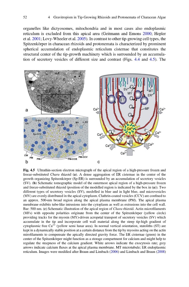
structural center of the tip-growth machinery which is surrounded by an accumulation of secretory vesicles of different size and contrast (Figs. 4.4 and 4.5). The
Fig. 4.5 Ultrathin-section electron micrograph of the apical region of a high-pressure frozen and
freeze-substituted Chara rhizoid (a). A dense aggregation of ER cisternae in the center of the
growth organizing Spitzenkörper (Sp-ER) is surrounded by an accumulation of secretory vesicles
(SV). (b) Schematic tomographic model of the outermost apical region of a high-pressure frozen
and freeze-substituted rhizoid (position of the modelled region is indicated by the box in (a)). Two
different types of secretory vesicles (SV), modelled in blue and in light blue, and microvesicles
(MV) are evenly distributed in the apical cytoplasm. Clathrin-coated vesicles (CCV) are confined to
an approx. 500-nm broad region along the apical plasma membrane (PM). The apical plasma
membrane exhibits tube-like intrusions into the cytoplasm as well as extrusions into the cell wall.
Bar: 500 nm. (c) Schematic illustration of the apical region of Chara rhizoid. Actin microfilaments
(MFs) with opposite polarities originate from the center of the Spitzenkörper (yellow circle)
providing tracks for the myosin (MY)-driven acropetal transport of secretory vesicles (SV) which
accumulate in the tip and incorporate cell wall material along the steep tip-high gradient of
cytoplasmic free Ca
2+ (yellow semi lunar area). In normal vertical orientation, statoliths (ST) are
kept in a dynamically stable position at a certain distance from the tip by myosins acting on the actin
mirofilaments to compensate the apically directed gravity force. The ER cisternae (green) in the
center of the Spitzenkörper might function as a storage compartment for calcium and might help to
regulate the steepness of the calcium gradient. White arrows indicate the exocytosis rate; grey
arrows indicate calcium fluxes at the apical plasma membrane. MT microtubule; ER endoplasmic
reticulum. Images were modified after Braun and Limbach (2006) and Limbach and Braun (2008)
52
4 Gravitropism in Tip-Growing Rhizoids and Protonemata of Characean Algae
reticulum is excluded from this apical area (Geitmann and Emons 2000; Hepler
et al. 2001; Lovy-Wheeler et al. 2005). In contrast to other tip-growing cell types, the
Spitzenkörper in characean rhizoids and protonemata is characterized by prominent
spherical accumulation of endoplasmic reticulum cisternae that constitutes the
structural center of the tip-growth machinery which is surrounded by an accumulation of secretory vesicles of different size and contrast (Figs. 4.4 and 4.5). The
Fig. 4.5 Ultrathin-section electron micrograph of the apical region of a high-pressure frozen and
freeze-substituted Chara rhizoid (a). A dense aggregation of ER cisternae in the center of the
growth organizing Spitzenkörper (Sp-ER) is surrounded by an accumulation of secretory vesicles
(SV). (b) Schematic tomographic model of the outermost apical region of a high-pressure frozen
and freeze-substituted rhizoid (position of the modelled region is indicated by the box in (a)). Two
different types of secretory vesicles (SV), modelled in blue and in light blue, and microvesicles
(MV) are evenly distributed in the apical cytoplasm. Clathrin-coated vesicles (CCV) are confined to
an approx. 500-nm broad region along the apical plasma membrane (PM). The apical plasma
membrane exhibits tube-like intrusions into the cytoplasm as well as extrusions into the cell wall.
Bar: 500 nm. (c) Schematic illustration of the apical region of Chara rhizoid. Actin microfilaments
(MFs) with opposite polarities originate from the center of the Spitzenkörper (yellow circle)
providing tracks for the myosin (MY)-driven acropetal transport of secretory vesicles (SV) which
accumulate in the tip and incorporate cell wall material along the steep tip-high gradient of
cytoplasmic free Ca
2+ (yellow semi lunar area). In normal vertical orientation, statoliths (ST) are
kept in a dynamically stable position at a certain distance from the tip by myosins acting on the actin
mirofilaments to compensate the apically directed gravity force. The ER cisternae (green) in the
center of the Spitzenkörper might function as a storage compartment for calcium and might help to
regulate the steepness of the calcium gradient. White arrows indicate the exocytosis rate; grey
arrows indicate calcium fluxes at the apical plasma membrane. MT microtubule; ER endoplasmic
reticulum. Images were modified after Braun and Limbach (2006) and Limbach and Braun (2008)
52
4 Gravitropism in Tip-Growing Rhizoids and Protonemata of Characean Algae