

65
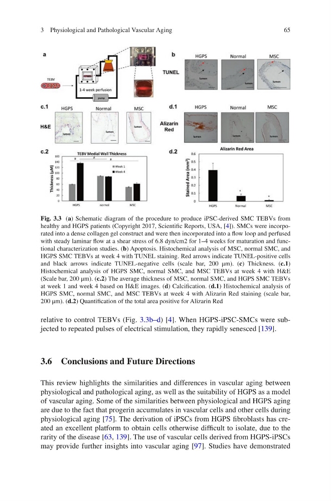
relative to control TEBVs (Fig. 3.3b–d) [4]. When HGPS-iPSC-SMCs were subjected to repeated pulses of electrical stimulation, they rapidly senesced [139].
3.6 Conclusions and Future Directions
This review highlights the similarities and differences in vascular aging between
physiological and pathological aging, as well as the suitability of HGPS as a model
of vascular aging. Some of the similarities between physiological and HGPS aging
are due to the fact that progerin accumulates in vascular cells and other cells during
physiological aging [75]. The derivation of iPSCs from HGPS fibroblasts has created an excellent platform to obtain cells otherwise difficult to isolate, due to the
rarity of the disease [63, 139]. The use of vascular cells derived from HGPS-iPSCs
may provide further insights into vascular aging [97]. Studies have demonstrated
Fig. 3.3 (a) Schematic diagram of the procedure to produce iPSC-derived SMC TEBVs from
healthy and HGPS patients (Copyright 2017, Scientific Reports, USA, [4]). SMCs were incorporated into a dense collagen gel construct and were then incorporated into a flow loop and perfused
with steady laminar flow at a shear stress of 6.8 dyn/cm2 for 1–4 weeks for maturation and functional characterization studies. (b) Apoptosis. Histochemical analysis of MSC, normal SMC, and
HGPS SMC TEBVs at week 4 with TUNEL staining. Red arrows indicate TUNEL-positive cells
and black arrows indicate TUNEL-negative cells (scale bar, 200 μm). (c) Thickness. (c.1)
Histochemical analysis of HGPS SMC, normal SMC, and MSC TEBVs at week 4 with H&E
(Scale bar, 200 μm). (c.2) The average thickness of MSC, normal SMC, and HGPS SMC TEBVs
at week 1 and week 4 based on H&E images. (d) Calcification. (d.1) Histochemical analysis of
HGPS SMC, normal SMC, and MSC TEBVs at week 4 with Alizarin Red staining (scale bar,
200 μm). (d.2) Quantification of the total area positive for Alizarin Red
3 Physiological and Pathological Vascular Aging
relative to control TEBVs (Fig. 3.3b–d) [4]. When HGPS-iPSC-SMCs were subjected to repeated pulses of electrical stimulation, they rapidly senesced [139].
3.6 Conclusions and Future Directions
This review highlights the similarities and differences in vascular aging between
physiological and pathological aging, as well as the suitability of HGPS as a model
of vascular aging. Some of the similarities between physiological and HGPS aging
are due to the fact that progerin accumulates in vascular cells and other cells during
physiological aging [75]. The derivation of iPSCs from HGPS fibroblasts has created an excellent platform to obtain cells otherwise difficult to isolate, due to the
rarity of the disease [63, 139]. The use of vascular cells derived from HGPS-iPSCs
may provide further insights into vascular aging [97]. Studies have demonstrated
Fig. 3.3 (a) Schematic diagram of the procedure to produce iPSC-derived SMC TEBVs from
healthy and HGPS patients (Copyright 2017, Scientific Reports, USA, [4]). SMCs were incorporated into a dense collagen gel construct and were then incorporated into a flow loop and perfused
with steady laminar flow at a shear stress of 6.8 dyn/cm2 for 1–4 weeks for maturation and functional characterization studies. (b) Apoptosis. Histochemical analysis of MSC, normal SMC, and
HGPS SMC TEBVs at week 4 with TUNEL staining. Red arrows indicate TUNEL-positive cells
and black arrows indicate TUNEL-negative cells (scale bar, 200 μm). (c) Thickness. (c.1)
Histochemical analysis of HGPS SMC, normal SMC, and MSC TEBVs at week 4 with H&E
(Scale bar, 200 μm). (c.2) The average thickness of MSC, normal SMC, and HGPS SMC TEBVs
at week 1 and week 4 based on H&E images. (d) Calcification. (d.1) Histochemical analysis of
HGPS SMC, normal SMC, and MSC TEBVs at week 4 with Alizarin Red staining (scale bar,
200 μm). (d.2) Quantification of the total area positive for Alizarin Red
3 Physiological and Pathological Vascular Aging