

148
Cells
Culture conditions
Cardiac characterization
Vascular characterization
Ref.
hiPSC-CM hiPSC-CM
• Two million cells
• Percentage: 33%
hiPSC-SMC
• Two million cells
• Percentage: 33%
hiPSC-EC
• Two million cells
• Percentage: 33%
• Fibrin hydrogel loaded with IGF-1 for cell
survival
• Directly injected cells into hydrogel on the
myocardium
• Cells are differentiated into three lineages
(CM, SMC, and EC) and injected directly into
hydrogel without in vitro culture
Advantages:
• Improved functionality in vivo with triculture and
patch present and reduced CM apoptosis of myocytes
• Increased ejection fraction
• Decreased infarct size
• Cell survival was greatest in tri-culture
cells + patch group
Limitations:
• Cardiac electrophysiology not characterized prior
to in vivo implantation
Advantages:
• Tri-culture promoted survival and
proliferation of cardiomyocytes
• Engineered vasculature promotes host
vasculature angiogenic response
Limitations:
• Vasculature was not characterized before
implantation into the infarcted myocardium
[134]
hiPSC-ECM
• One million cells
• Percentage: 76%
hMSC
• 400,000 cells (w/
hiPSC-ECM)
• 300,000 cells (w/hCMVECs)
• Percentage: 23%/38%
hCMVECs
• 500,000 cells
• Percentage: 63%
• 3D collagen cell carrier
• hMSC/hCMVECs culture for 7 days under
vascular conditions
• hMSC/hiPSC-ECMs cultured for 7–14 days on
top pre vascularized cultures
Advantages:
• Positive staining for cardiac MHC across the
construct
• Rhythmic contractions
• Calcium flux monitored across system
`Limitations:
• Cardiomyocyte orientation not controlled
• Electrophysiological parameters not examined
Advantages:
• CD31 positive cord-like structures
established
• Pericyte-like cell stained positive for SMA
• Sequential seeding of vasculature cells
• Positive staining for AcLDL and vWF in
the tri-culture system
Limitations:
• Vasculature is reduced in the presence of
all three cell types
[122]
hiPSC-CM
• 14,850–23,100 cells
• Percentage: 45–70%
hCF
• 0 to 13,200 cells
• Percentage: 0–40%
HUVECs
• 4950–9900 cells
• Percentage: 15–30%
• 3D bioprinting (0.32 cm
3
)
• Single layer of cell aggregates (cardiospheres)
that were 450–550 μm in diameter
• Printed on needle Array
• Cardiac patch was matured for 72 h with
needle array in place
• Cardiospheres beat after 2 days and fused after
removal of needle array
• Cardiospheres were implanted onto
myocardium and secured with tissue glue in vivo
Advantages:
• Electrophysiological parameters quantified
• Ventricular-like action potentials
• Developed gap junctions (Cx-43) to propagate
signal as a syncytium
• Low levels of cell death from TUNEL assay
• Conduction velocity near 4.6 cm/s
Limitations:
• Conduction velocity decreased with increased
fibroblast percentage
Advantages:
• CD31 staining throughout the cardiosphere
• Nascent vasculature development in vivo
Limitations:
• Vasculature not quantified
• Presence of 3D lumens not investigate or
represented
[85]
hiPSC-CM
• 4 M/mL
• Percentage: 65%
hPC
• 0.36 M/mL
• Percentage: 6%
BOEC
• 1.82 M/mL cells
• Percentage: 29%
• Fibrin hydrogel
• 8.4 mm × 5 mm
• 4 mg/mL fibrinogen
• 120 μL total volume
• Bilayer patch was created by coating with
fibrinogen and 10 U/mL thrombin
• Grown for 14 days
Advantages:
• Bilayer patch resulted in increased CM density
compared to control (237 vs 35 CM/mm
2
)
• CM only patches underwent more apoptosis than
bilayer (75.4% vs 37.5%)
• Bilayer patch output was 2.17 nN/input CM vs
0.3 nN/input CM
Limitations:
• Electrophysiological properties not analyzed
• Alignment of cardiomyocyte
Advantages:
• ~34 lumens/mm
2
• ~65 vessels/mm
2
in vivo
• ~67% perfused fraction of vasculature
Limitations:
• Vasculature developed independently of
cardiomyocyte culture which is not similar
to the native myocardium
[101]
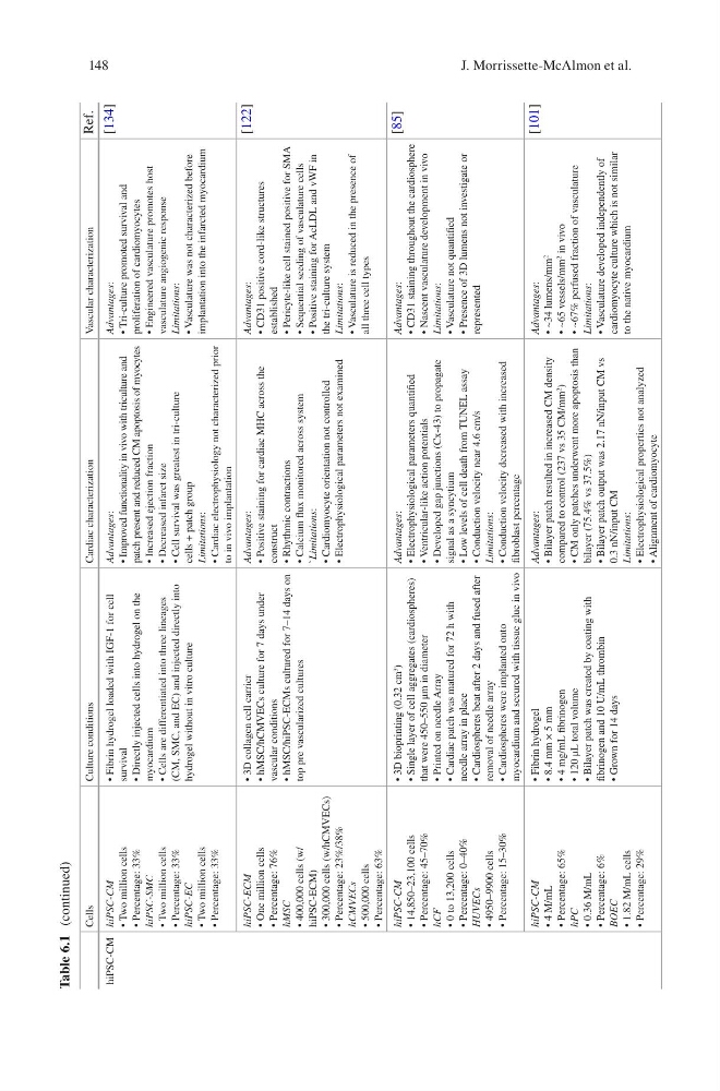
Table 6.1 (continued)
J. Morrissette-McAlmon et al.
Cells
Culture conditions
Cardiac characterization
Vascular characterization
Ref.
hiPSC-CM hiPSC-CM
• Two million cells
• Percentage: 33%
hiPSC-SMC
• Two million cells
• Percentage: 33%
hiPSC-EC
• Two million cells
• Percentage: 33%
• Fibrin hydrogel loaded with IGF-1 for cell
survival
• Directly injected cells into hydrogel on the
myocardium
• Cells are differentiated into three lineages
(CM, SMC, and EC) and injected directly into
hydrogel without in vitro culture
Advantages:
• Improved functionality in vivo with triculture and
patch present and reduced CM apoptosis of myocytes
• Increased ejection fraction
• Decreased infarct size
• Cell survival was greatest in tri-culture
cells + patch group
Limitations:
• Cardiac electrophysiology not characterized prior
to in vivo implantation
Advantages:
• Tri-culture promoted survival and
proliferation of cardiomyocytes
• Engineered vasculature promotes host
vasculature angiogenic response
Limitations:
• Vasculature was not characterized before
implantation into the infarcted myocardium
[134]
hiPSC-ECM
• One million cells
• Percentage: 76%
hMSC
• 400,000 cells (w/
hiPSC-ECM)
• 300,000 cells (w/hCMVECs)
• Percentage: 23%/38%
hCMVECs
• 500,000 cells
• Percentage: 63%
• 3D collagen cell carrier
• hMSC/hCMVECs culture for 7 days under
vascular conditions
• hMSC/hiPSC-ECMs cultured for 7–14 days on
top pre vascularized cultures
Advantages:
• Positive staining for cardiac MHC across the
construct
• Rhythmic contractions
• Calcium flux monitored across system
`Limitations:
• Cardiomyocyte orientation not controlled
• Electrophysiological parameters not examined
Advantages:
• CD31 positive cord-like structures
established
• Pericyte-like cell stained positive for SMA
• Sequential seeding of vasculature cells
• Positive staining for AcLDL and vWF in
the tri-culture system
Limitations:
• Vasculature is reduced in the presence of
all three cell types
[122]
hiPSC-CM
• 14,850–23,100 cells
• Percentage: 45–70%
hCF
• 0 to 13,200 cells
• Percentage: 0–40%
HUVECs
• 4950–9900 cells
• Percentage: 15–30%
• 3D bioprinting (0.32 cm
3
)
• Single layer of cell aggregates (cardiospheres)
that were 450–550 μm in diameter
• Printed on needle Array
• Cardiac patch was matured for 72 h with
needle array in place
• Cardiospheres beat after 2 days and fused after
removal of needle array
• Cardiospheres were implanted onto
myocardium and secured with tissue glue in vivo
Advantages:
• Electrophysiological parameters quantified
• Ventricular-like action potentials
• Developed gap junctions (Cx-43) to propagate
signal as a syncytium
• Low levels of cell death from TUNEL assay
• Conduction velocity near 4.6 cm/s
Limitations:
• Conduction velocity decreased with increased
fibroblast percentage
Advantages:
• CD31 staining throughout the cardiosphere
• Nascent vasculature development in vivo
Limitations:
• Vasculature not quantified
• Presence of 3D lumens not investigate or
represented
[85]
hiPSC-CM
• 4 M/mL
• Percentage: 65%
hPC
• 0.36 M/mL
• Percentage: 6%
BOEC
• 1.82 M/mL cells
• Percentage: 29%
• Fibrin hydrogel
• 8.4 mm × 5 mm
• 4 mg/mL fibrinogen
• 120 μL total volume
• Bilayer patch was created by coating with
fibrinogen and 10 U/mL thrombin
• Grown for 14 days
Advantages:
• Bilayer patch resulted in increased CM density
compared to control (237 vs 35 CM/mm
2
)
• CM only patches underwent more apoptosis than
bilayer (75.4% vs 37.5%)
• Bilayer patch output was 2.17 nN/input CM vs
0.3 nN/input CM
Limitations:
• Electrophysiological properties not analyzed
• Alignment of cardiomyocyte
Advantages:
• ~34 lumens/mm
2
• ~65 vessels/mm
2
in vivo
• ~67% perfused fraction of vasculature
Limitations:
• Vasculature developed independently of
cardiomyocyte culture which is not similar
to the native myocardium
[101]
Table 6.1 (continued)
J. Morrissette-McAlmon et al.