

143
6.2 Tissue-Engineered Cardiac Patches
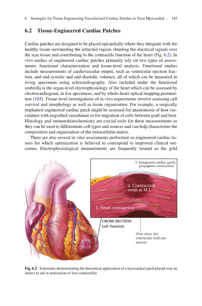
Cardiac patches are designed to be placed epicardially where they integrate with the
healthy tissue surrounding the infarcted region, shunting the electrical signals over
the scar tissue and contributing to the contractile function of the heart (Fig. 6.2). In
vivo studies of engineered cardiac patches primarily rely on two types of assessments: functional characterization and tissue-level analysis. Functional studies
include measurements of cardiovascular output, such as ventricular ejection fraction, and end-systolic and end-diastolic volumes, all of which can be measured in
living specimens using echocardiography. Also included under the functional
umbrella is the organ-level electrophysiology of the heart which can be assessed by
electrocardiogram, in live specimens, and by whole-heart optical mapping postmortem [103]. Tissue-level investigations of in vivo experiments involve assessing cell
survival and morphology as well as tissue organization. For example, a surgically
implanted engineered cardiac patch might be assessed for anastomosis of host vasculature with engrafted vasculature or for migration of cells between graft and host.
Histology and immunohistochemistry are crucial tools for these measurements as
they can be used to differentiate cell types and sources and can help characterize the
composition and organization of the extracellular matrix.
There are also several in vitro assessments performed on engineered cardiac tissues for which optimization is believed to correspond to improved clinical outcomes. Electrophysiological measurements are frequently treated as the gold
Fig. 6.2 Schematic demonstrating the theoretical application of a myocardial patch placed over an
infarct to aid in restoration of lost contractility
6 Strategies for Tissue Engineering Vascularized Cardiac Patches to Treat Myocardial…
6.2 Tissue-Engineered Cardiac Patches
Cardiac patches are designed to be placed epicardially where they integrate with the
healthy tissue surrounding the infarcted region, shunting the electrical signals over
the scar tissue and contributing to the contractile function of the heart (Fig. 6.2). In
vivo studies of engineered cardiac patches primarily rely on two types of assessments: functional characterization and tissue-level analysis. Functional studies
include measurements of cardiovascular output, such as ventricular ejection fraction, and end-systolic and end-diastolic volumes, all of which can be measured in
living specimens using echocardiography. Also included under the functional
umbrella is the organ-level electrophysiology of the heart which can be assessed by
electrocardiogram, in live specimens, and by whole-heart optical mapping postmortem [103]. Tissue-level investigations of in vivo experiments involve assessing cell
survival and morphology as well as tissue organization. For example, a surgically
implanted engineered cardiac patch might be assessed for anastomosis of host vasculature with engrafted vasculature or for migration of cells between graft and host.
Histology and immunohistochemistry are crucial tools for these measurements as
they can be used to differentiate cell types and sources and can help characterize the
composition and organization of the extracellular matrix.
There are also several in vitro assessments performed on engineered cardiac tissues for which optimization is believed to correspond to improved clinical outcomes. Electrophysiological measurements are frequently treated as the gold
Fig. 6.2 Schematic demonstrating the theoretical application of a myocardial patch placed over an
infarct to aid in restoration of lost contractility
6 Strategies for Tissue Engineering Vascularized Cardiac Patches to Treat Myocardial…