

142
include stem cell transplantation, thrombolytic therapy, and stenting of coronary
arteries, have improved myocardial salvage, but therapies that directly induce regeneration of the ventricular wall and improvement in functionality are lacking.
Cardiovascular tissue engineering has the potential to ameliorate the damage
resulting from the limited endogenous repair mechanisms of the heart following MI
[45]. Currently, there are no methods that completely repair the damaged myocardium post injury. Tissue engineering approaches aim to regenerate the myocardium
by mimicking the tissue structure and cell composition. The native makeup of the
myocardium includes a host of cells that are critical for survival, maturation, and
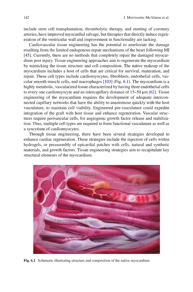
repair. These cell types include cardiomyocytes, fibroblasts, endothelial cells, vascular smooth muscle cells, and macrophages [103] (Fig. 6.1). The myocardium is a
highly metabolic, vascularized tissue characterized by having three endothelial cells
to every one cardiomyocyte and an intercapillary distance of 15–50 μm [62]. Tissue
engineering of the myocardium requires the development of adequate interconnected capillary networks that have the ability to anastomose quickly with the host
vasculature, to maintain cell viability. Engineered pre-vasculature could expedite
integration of the graft with host tissue and enhance regeneration. Vascular structures require perivascular cells, for angiogenic growth factor release and stabilization. Thus, multiple cell types are required to form functional vasculature as well as
a syncytium of cardiomyocytes.
Through tissue engineering, there have been several strategies developed to
enhance cardiac regeneration. These strategies include the injection of cells within
hydrogels, or preassembly of epicardial patches with cells, natural and synthetic
materials, and growth factors. Tissue engineering strategies aim to recapitulate key
structural elements of the myocardium.
Fig. 6.1 Schematic illustrating structure and composition of the native myocardium
J. Morrissette-McAlmon et al.
include stem cell transplantation, thrombolytic therapy, and stenting of coronary
arteries, have improved myocardial salvage, but therapies that directly induce regeneration of the ventricular wall and improvement in functionality are lacking.
Cardiovascular tissue engineering has the potential to ameliorate the damage
resulting from the limited endogenous repair mechanisms of the heart following MI
[45]. Currently, there are no methods that completely repair the damaged myocardium post injury. Tissue engineering approaches aim to regenerate the myocardium
by mimicking the tissue structure and cell composition. The native makeup of the
myocardium includes a host of cells that are critical for survival, maturation, and
repair. These cell types include cardiomyocytes, fibroblasts, endothelial cells, vascular smooth muscle cells, and macrophages [103] (Fig. 6.1). The myocardium is a
highly metabolic, vascularized tissue characterized by having three endothelial cells
to every one cardiomyocyte and an intercapillary distance of 15–50 μm [62]. Tissue
engineering of the myocardium requires the development of adequate interconnected capillary networks that have the ability to anastomose quickly with the host
vasculature, to maintain cell viability. Engineered pre-vasculature could expedite
integration of the graft with host tissue and enhance regeneration. Vascular structures require perivascular cells, for angiogenic growth factor release and stabilization. Thus, multiple cell types are required to form functional vasculature as well as
a syncytium of cardiomyocytes.
Through tissue engineering, there have been several strategies developed to
enhance cardiac regeneration. These strategies include the injection of cells within
hydrogels, or preassembly of epicardial patches with cells, natural and synthetic
materials, and growth factors. Tissue engineering strategies aim to recapitulate key
structural elements of the myocardium.
Fig. 6.1 Schematic illustrating structure and composition of the native myocardium
J. Morrissette-McAlmon et al.