

128
5.2.2.2 Application
Microvascular Network Creation (Table 5.1)
Khalil et al. used a pneumatic nozzle system with an aqueous alginate solution and
rat heart endothelial cells (RHECs), to print a microvascular network [27]. The
group demonstrated a high live/dead RHEC cell ratio in the printed vascular scaffold, even 14 days after printing. Furthermore, the mechanical properties of the
printed scaffold were unchanged over 3 weeks post-printing. In order to evaluate the
capillary system construction, a built-in microchannel system within the vascular
constructs was developed by Gao et al. [15]. A coaxial nozzle system, with an inner
flow of calcium chloride and outer flow of alginate solution, was used. These microchannels formed the interface between calcium chloride and sodium alginate solutions, where the cross-linkage took place. The group described the creation of
parallel microchannels alternating in two different sizes of channel, using the additional ungelled sodium alginate for hollow filament deposition. The ungelled
sodium alginate was deposited on the outer surface of each individual channel, so as
to form a parallel network. Then hollow channels, in between the regular channels,
were created by removal of the ungelled printed alginate. The group also demonstrated improved cell viability using vascular constructs with microchannels, versus
those without microchannels, when the sodium alginate scaffolds were co-printed
with mouse fibroblasts.
The microvascular system was further investigated by Colosi et al. [3] using a
coaxial nozzle extrusion system to print a microfluidic channel composed of low
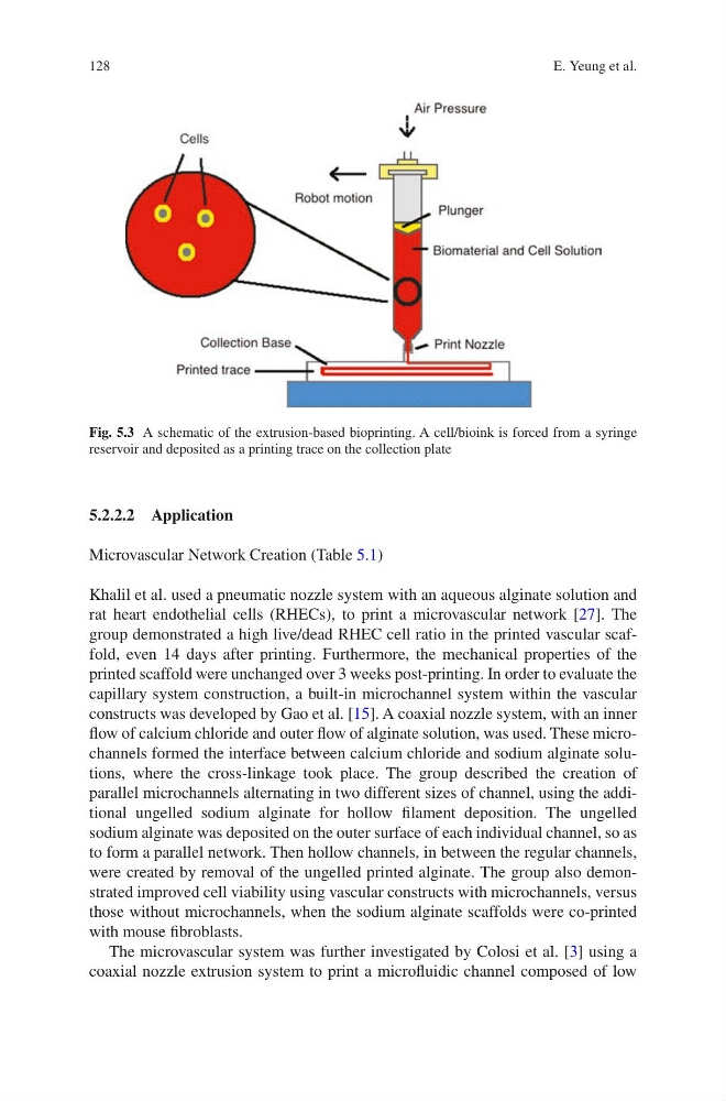
Fig. 5.3 A schematic of the extrusion-based bioprinting. A cell/bioink is forced from a syringe
reservoir and deposited as a printing trace on the collection plate
E. Yeung et al.
5.2.2.2 Application
Microvascular Network Creation (Table 5.1)
Khalil et al. used a pneumatic nozzle system with an aqueous alginate solution and
rat heart endothelial cells (RHECs), to print a microvascular network [27]. The
group demonstrated a high live/dead RHEC cell ratio in the printed vascular scaffold, even 14 days after printing. Furthermore, the mechanical properties of the
printed scaffold were unchanged over 3 weeks post-printing. In order to evaluate the
capillary system construction, a built-in microchannel system within the vascular
constructs was developed by Gao et al. [15]. A coaxial nozzle system, with an inner
flow of calcium chloride and outer flow of alginate solution, was used. These microchannels formed the interface between calcium chloride and sodium alginate solutions, where the cross-linkage took place. The group described the creation of
parallel microchannels alternating in two different sizes of channel, using the additional ungelled sodium alginate for hollow filament deposition. The ungelled
sodium alginate was deposited on the outer surface of each individual channel, so as
to form a parallel network. Then hollow channels, in between the regular channels,
were created by removal of the ungelled printed alginate. The group also demonstrated improved cell viability using vascular constructs with microchannels, versus
those without microchannels, when the sodium alginate scaffolds were co-printed
with mouse fibroblasts.
The microvascular system was further investigated by Colosi et al. [3] using a
coaxial nozzle extrusion system to print a microfluidic channel composed of low
Fig. 5.3 A schematic of the extrusion-based bioprinting. A cell/bioink is forced from a syringe
reservoir and deposited as a printing trace on the collection plate
E. Yeung et al.