

127
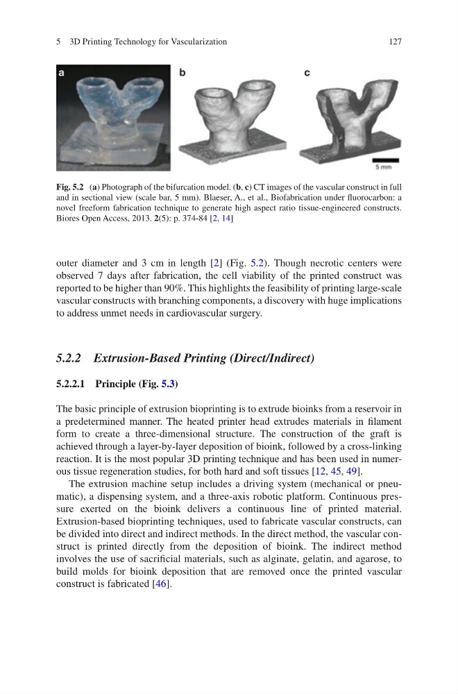
outer diameter and 3 cm in length [2] (Fig. 5.2). Though necrotic centers were
observed 7 days after fabrication, the cell viability of the printed construct was
reported to be higher than 90%. This highlights the feasibility of printing large-scale
vascular constructs with branching components, a discovery with huge implications
to address unmet needs in cardiovascular surgery.
5.2.2 Extrusion-Based Printing (Direct/Indirect)
5.2.2.1 Principle (Fig. 5.3)
The basic principle of extrusion bioprinting is to extrude bioinks from a reservoir in
a predetermined manner. The heated printer head extrudes materials in filament
form to create a three-dimensional structure. The construction of the graft is
achieved through a layer-by-layer deposition of bioink, followed by a cross-linking
reaction. It is the most popular 3D printing technique and has been used in numerous tissue regeneration studies, for both hard and soft tissues [12, 45, 49].
The extrusion machine setup includes a driving system (mechanical or pneumatic), a dispensing system, and a three-axis robotic platform. Continuous pressure exerted on the bioink delivers a continuous line of printed material.
Extrusion-based bioprinting techniques, used to fabricate vascular constructs, can
be divided into direct and indirect methods. In the direct method, the vascular construct is printed directly from the deposition of bioink. The indirect method
involves the use of sacrificial materials, such as alginate, gelatin, and agarose, to
build molds for bioink deposition that are removed once the printed vascular
construct is fabricated [46].
Fig. 5.2 (a) Photograph of the bifurcation model. (b, c) CT images of the vascular construct in full
and in sectional view (scale bar, 5 mm). Blaeser, A., et al., Biofabrication under fluorocarbon: a
novel freeform fabrication technique to generate high aspect ratio tissue-engineered constructs.
Biores Open Access, 2013. 2(5): p. 374-84 [2, 14]
5 3D Printing Technology for Vascularization
outer diameter and 3 cm in length [2] (Fig. 5.2). Though necrotic centers were
observed 7 days after fabrication, the cell viability of the printed construct was
reported to be higher than 90%. This highlights the feasibility of printing large-scale
vascular constructs with branching components, a discovery with huge implications
to address unmet needs in cardiovascular surgery.
5.2.2 Extrusion-Based Printing (Direct/Indirect)
5.2.2.1 Principle (Fig. 5.3)
The basic principle of extrusion bioprinting is to extrude bioinks from a reservoir in
a predetermined manner. The heated printer head extrudes materials in filament
form to create a three-dimensional structure. The construction of the graft is
achieved through a layer-by-layer deposition of bioink, followed by a cross-linking
reaction. It is the most popular 3D printing technique and has been used in numerous tissue regeneration studies, for both hard and soft tissues [12, 45, 49].
The extrusion machine setup includes a driving system (mechanical or pneumatic), a dispensing system, and a three-axis robotic platform. Continuous pressure exerted on the bioink delivers a continuous line of printed material.
Extrusion-based bioprinting techniques, used to fabricate vascular constructs, can
be divided into direct and indirect methods. In the direct method, the vascular construct is printed directly from the deposition of bioink. The indirect method
involves the use of sacrificial materials, such as alginate, gelatin, and agarose, to
build molds for bioink deposition that are removed once the printed vascular
construct is fabricated [46].
Fig. 5.2 (a) Photograph of the bifurcation model. (b, c) CT images of the vascular construct in full
and in sectional view (scale bar, 5 mm). Blaeser, A., et al., Biofabrication under fluorocarbon: a
novel freeform fabrication technique to generate high aspect ratio tissue-engineered constructs.
Biores Open Access, 2013. 2(5): p. 374-84 [2, 14]
5 3D Printing Technology for Vascularization