

125
5.2.1.2 Application
Microvascular Network Creation (Table 5.1)
Nishiyama et al. printed vascular constructs following the same method described
above, using a custom inkjet bioprinter with a position detection accuracy of less
than a few micrometers [39]. This experiment demonstrated a technique for bioprinting a 3D structure using a laminating method, which involves the sequential
deposition of materials. They used a viscosity enhancer to consolidate and fortify
the constructs. The group further investigated the use of such technology to fabricate smaller-scale tubular constructs (200 um in diameter, compared to the original
1 mm diameter tubes), using a small-sized nozzle head, to downsize the printed
hydrogel droplets from 40 um to 25 um [40]. These smaller-sized tubular structures
signified the potential for the use of this technology in vascular tissue engineering.
Cui et al. used bioink in a modified HP Deskjet 600 thermal printer to print
microvascular constructs. Human microvascular endothelial cells (HMVECs) in a
fibrin hydrogel were concurrently deposited into a thrombin substrate [4]. In
3 weeks, printed microvasculature lined by HMVEC was observed in the scaffold.
Similarly, Zhao et al. developed a perfusable vascular channel using rat collagen
type 1, human umbilical vein endothelial cells (HUVECs), and gelatin, to construct
a vascular network [55]. Mesoscopic fluorescence molecular tomography (MFMT)
demonstrated that the endothelial cells, which lined in the inner layer of the branching vascular networks, were evenly distributed and showed signs of migration in the
luminal surface. Further, this experiment also provided a visualization of fluid flow
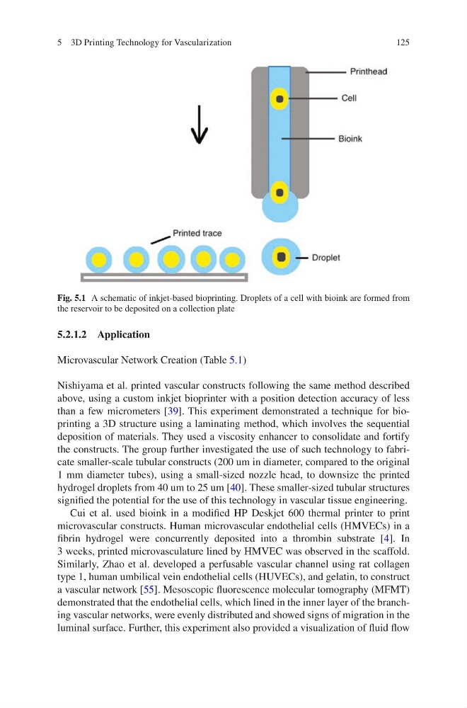
Fig. 5.1 A schematic of inkjet-based bioprinting. Droplets of a cell with bioink are formed from
the reservoir to be deposited on a collection plate
5 3D Printing Technology for Vascularization
5.2.1.2 Application
Microvascular Network Creation (Table 5.1)
Nishiyama et al. printed vascular constructs following the same method described
above, using a custom inkjet bioprinter with a position detection accuracy of less
than a few micrometers [39]. This experiment demonstrated a technique for bioprinting a 3D structure using a laminating method, which involves the sequential
deposition of materials. They used a viscosity enhancer to consolidate and fortify
the constructs. The group further investigated the use of such technology to fabricate smaller-scale tubular constructs (200 um in diameter, compared to the original
1 mm diameter tubes), using a small-sized nozzle head, to downsize the printed
hydrogel droplets from 40 um to 25 um [40]. These smaller-sized tubular structures
signified the potential for the use of this technology in vascular tissue engineering.
Cui et al. used bioink in a modified HP Deskjet 600 thermal printer to print
microvascular constructs. Human microvascular endothelial cells (HMVECs) in a
fibrin hydrogel were concurrently deposited into a thrombin substrate [4]. In
3 weeks, printed microvasculature lined by HMVEC was observed in the scaffold.
Similarly, Zhao et al. developed a perfusable vascular channel using rat collagen
type 1, human umbilical vein endothelial cells (HUVECs), and gelatin, to construct
a vascular network [55]. Mesoscopic fluorescence molecular tomography (MFMT)
demonstrated that the endothelial cells, which lined in the inner layer of the branching vascular networks, were evenly distributed and showed signs of migration in the
luminal surface. Further, this experiment also provided a visualization of fluid flow
Fig. 5.1 A schematic of inkjet-based bioprinting. Droplets of a cell with bioink are formed from
the reservoir to be deposited on a collection plate
5 3D Printing Technology for Vascularization