

95
In addition to the effects of matrix stiffness on postnatal vascular regeneration,
matrix stiffness has been probed as an important regulator of stem cell fate.
Beginning with the pioneering work of Engler et al. [60], studies examining the
effects of substrate stiffness and mechanical signaling transduction pathways on
stem cell fate have proven instrumental in enhancing our collective knowledge of
differentiation schema. To this end, our group has shown that substrate stiffness can
govern EC fate through alterations in mesodermal precursors. Similar to the
enhancements in EC fate observed upon culture in low O 2 environments (Fig. 4.5a–
d) [135], compliant substrates enhance mesodermal differentiation, which results in
robust EC differentiation (Fig. 4.5e–i) [205].
A recent illuminating study identified stress relaxation as an important, yet
understudied, regulator of mechanical signal transduction. Specifically, in
alginate- based hydrogels with the same matrix stiffness and pore size, altering stress
relaxation modulated MSC cell fate [34]. While studies of the effect of stress relaxation on EC fate and vascular morphogenesis have not been published, stress relaxation is an important parameter to bear in mind for biomaterial design, particularly
because covalently cross-linked hydrogels do not exhibit stress relaxation behavior
similar to that of the native ECM.
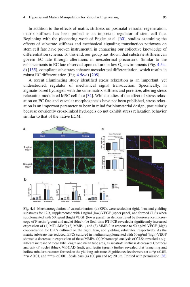
Fig. 4.4 Mechanoregulation of vascularization. (a) EPCs were seeded on rigid, firm, and yielding
substrates for 12 h, supplemented with 1 ng/ml (low) VEGF (upper panel) and formed CLSs when
supplemented with 50 ng/ml (high) VEGF (lower panel), as demonstrated by fluorescence microscopy of F-actin (green) and nuclei (blue). (b) Real-time RT-PCR revealed a significantly increased
expression of (1) MT1-MMP, (2) MMP-1, and (3) MMP-2 in response to 50 ng/ml VEGF (high)
concentration for EPCs cultured on the rigid, firm, and yielding substrates, respectively. As the
matrix substrate was reduced, EPCs cultured in medium supplemented with 50 ng/ml (high) VEGF
showed a decrease in expression of these MMPs. (c) Metamorph analysis of CLSs revealed a significant increase of mean tube length and mean tube area, as substrate stiffness decreased. Confocal
analysis of nuclei (blue), VE-CAD (red), and lectin (green) further revealed that branching and
hollow tubular structures formed on the yielding substrate. Significance levels were set at *p < 0.05,
**p < 0.01, and ***p < 0.001. Scale bars (a) 100 μm and (c) 20 μm. Printed with permission [88]
4 Hypoxia and Matrix Manipulation for Vascular Engineering
In addition to the effects of matrix stiffness on postnatal vascular regeneration,
matrix stiffness has been probed as an important regulator of stem cell fate.
Beginning with the pioneering work of Engler et al. [60], studies examining the
effects of substrate stiffness and mechanical signaling transduction pathways on
stem cell fate have proven instrumental in enhancing our collective knowledge of
differentiation schema. To this end, our group has shown that substrate stiffness can
govern EC fate through alterations in mesodermal precursors. Similar to the
enhancements in EC fate observed upon culture in low O 2 environments (Fig. 4.5a–
d) [135], compliant substrates enhance mesodermal differentiation, which results in
robust EC differentiation (Fig. 4.5e–i) [205].
A recent illuminating study identified stress relaxation as an important, yet
understudied, regulator of mechanical signal transduction. Specifically, in
alginate- based hydrogels with the same matrix stiffness and pore size, altering stress
relaxation modulated MSC cell fate [34]. While studies of the effect of stress relaxation on EC fate and vascular morphogenesis have not been published, stress relaxation is an important parameter to bear in mind for biomaterial design, particularly
because covalently cross-linked hydrogels do not exhibit stress relaxation behavior
similar to that of the native ECM.
Fig. 4.4 Mechanoregulation of vascularization. (a) EPCs were seeded on rigid, firm, and yielding
substrates for 12 h, supplemented with 1 ng/ml (low) VEGF (upper panel) and formed CLSs when
supplemented with 50 ng/ml (high) VEGF (lower panel), as demonstrated by fluorescence microscopy of F-actin (green) and nuclei (blue). (b) Real-time RT-PCR revealed a significantly increased
expression of (1) MT1-MMP, (2) MMP-1, and (3) MMP-2 in response to 50 ng/ml VEGF (high)
concentration for EPCs cultured on the rigid, firm, and yielding substrates, respectively. As the
matrix substrate was reduced, EPCs cultured in medium supplemented with 50 ng/ml (high) VEGF
showed a decrease in expression of these MMPs. (c) Metamorph analysis of CLSs revealed a significant increase of mean tube length and mean tube area, as substrate stiffness decreased. Confocal
analysis of nuclei (blue), VE-CAD (red), and lectin (green) further revealed that branching and
hollow tubular structures formed on the yielding substrate. Significance levels were set at *p < 0.05,
**p < 0.01, and ***p < 0.001. Scale bars (a) 100 μm and (c) 20 μm. Printed with permission [88]
4 Hypoxia and Matrix Manipulation for Vascular Engineering