

2.3.5 CSTET and Elemental Analysis of Eukaryotic Cells
Eukaryotic cells have been studied extensively by CET tomography (recent
reviews: [85–87]), primarily at the very thin edges. Cell adhesion and cytoskeleton
structure can often be studied in detail. In order to access thicker regions, it is
necessary to produce thin lamellae by cryo-sectioning, either by the CEMOVIS
technique [88–90], or cryo-Focused Ion Beam (FIB) milling [91–93]. These
methods can produce spectacular results but the methodology for producing the thin
sections is highly challenging. In addition, the lamellae are removed from their
whole-cell context. By just being able to access three-fold thicker regions of
spreading vitrified cells, CSTET allows for access to far more cellular processes and
organelles, as schematically illustrated in Fig. 2.8.
Results with CSTET on eukaryotic cells have shown excellent visibility in the
reconstructions, even for regions as thick as one micron. In contrast to bacteria,
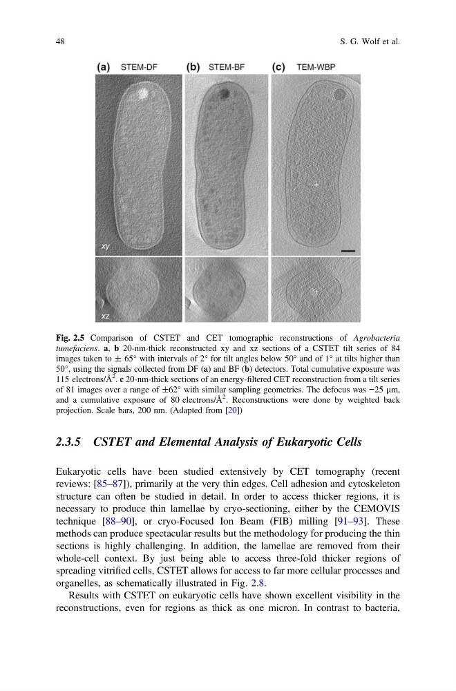
Fig. 2.5 Comparison of CSTET and CET tomographic reconstructions of Agrobacteria
tumefaciens. a, b 20-nm-thick reconstructed xy and xz sections of a CSTET tilt series of 84
images taken to ± 65° with intervals of 2° for tilt angles below 50° and of 1° at tilts higher than
50°, using the signals collected from DF (a) and BF (b) detectors. Total cumulative exposure was
115 electrons/Å
2
. c 20-nm-thick sections of an energy-filtered CET reconstruction from a tilt series
of 81 images over a range of ±62° with similar sampling geometries. The defocus was −25 lm,
and a cumulative exposure of 80 electrons/Å
2
. Reconstructions were done by weighted back
projection. Scale bars, 200 nm. (Adapted from [20])
48
S. G. Wolf et al.
Eukaryotic cells have been studied extensively by CET tomography (recent
reviews: [85–87]), primarily at the very thin edges. Cell adhesion and cytoskeleton
structure can often be studied in detail. In order to access thicker regions, it is
necessary to produce thin lamellae by cryo-sectioning, either by the CEMOVIS
technique [88–90], or cryo-Focused Ion Beam (FIB) milling [91–93]. These
methods can produce spectacular results but the methodology for producing the thin
sections is highly challenging. In addition, the lamellae are removed from their
whole-cell context. By just being able to access three-fold thicker regions of
spreading vitrified cells, CSTET allows for access to far more cellular processes and
organelles, as schematically illustrated in Fig. 2.8.
Results with CSTET on eukaryotic cells have shown excellent visibility in the
reconstructions, even for regions as thick as one micron. In contrast to bacteria,
Fig. 2.5 Comparison of CSTET and CET tomographic reconstructions of Agrobacteria
tumefaciens. a, b 20-nm-thick reconstructed xy and xz sections of a CSTET tilt series of 84
images taken to ± 65° with intervals of 2° for tilt angles below 50° and of 1° at tilts higher than
50°, using the signals collected from DF (a) and BF (b) detectors. Total cumulative exposure was
115 electrons/Å
2
. c 20-nm-thick sections of an energy-filtered CET reconstruction from a tilt series
of 81 images over a range of ±62° with similar sampling geometries. The defocus was −25 lm,
and a cumulative exposure of 80 electrons/Å
2
. Reconstructions were done by weighted back
projection. Scale bars, 200 nm. (Adapted from [20])
48
S. G. Wolf et al.