

1.10.3 Subtomogram averaging
Resolution of 4 nm must be improved upon to realise the full potential of molecular
cartography, and to decipher molecular function. The best resolution that can be
expected in electron tomography raw data, i.e. without interpolation artefacts and
signal loss at the detector, might never surpass 2 nm [109]; however, this would
suffice for the unambiguous structural determination of many intermediate-sized
molecular complexes. The improvements required for this are now being realised
via the use of phase plates [110] and direct electron detectors that account for all
electrons incident on the detector [111]. Considerable information is present beyond
(2 nm)
−1 , buried in noise [52]. This information can only be extracted using filtering techniques in conjunction with a priori knowledge of expected structure.
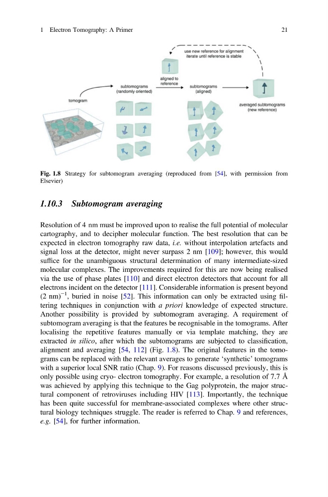
Another possibility is provided by subtomogram averaging. A requirement of
subtomogram averaging is that the features be recognisable in the tomograms. After
localising the repetitive features manually or via template matching, they are
extracted in silico, after which the subtomograms are subjected to classification,
alignment and averaging [54, 112] (Fig. 1.8). The original features in the tomograms can be replaced with the relevant averages to generate ‘synthetic’ tomograms
with a superior local SNR ratio (Chap. 9). For reasons discussed previously, this is
only possible using cryo- electron tomography. For example, a resolution of 7.7 Å
was achieved by applying this technique to the Gag polyprotein, the major structural component of retroviruses including HIV [113]. Importantly, the technique
has been quite successful for membrane-associated complexes where other structural biology techniques struggle. The reader is referred to Chap. 9 and references,
e.g. [54], for further information.
Fig. 1.8 Strategy for subtomogram averaging (reproduced from [54], with permission from
Elsevier)
1 Electron Tomography: A Primer
21
Resolution of 4 nm must be improved upon to realise the full potential of molecular
cartography, and to decipher molecular function. The best resolution that can be
expected in electron tomography raw data, i.e. without interpolation artefacts and
signal loss at the detector, might never surpass 2 nm [109]; however, this would
suffice for the unambiguous structural determination of many intermediate-sized
molecular complexes. The improvements required for this are now being realised
via the use of phase plates [110] and direct electron detectors that account for all
electrons incident on the detector [111]. Considerable information is present beyond
(2 nm)
−1 , buried in noise [52]. This information can only be extracted using filtering techniques in conjunction with a priori knowledge of expected structure.
Another possibility is provided by subtomogram averaging. A requirement of
subtomogram averaging is that the features be recognisable in the tomograms. After
localising the repetitive features manually or via template matching, they are
extracted in silico, after which the subtomograms are subjected to classification,
alignment and averaging [54, 112] (Fig. 1.8). The original features in the tomograms can be replaced with the relevant averages to generate ‘synthetic’ tomograms
with a superior local SNR ratio (Chap. 9). For reasons discussed previously, this is
only possible using cryo- electron tomography. For example, a resolution of 7.7 Å
was achieved by applying this technique to the Gag polyprotein, the major structural component of retroviruses including HIV [113]. Importantly, the technique
has been quite successful for membrane-associated complexes where other structural biology techniques struggle. The reader is referred to Chap. 9 and references,
e.g. [54], for further information.
Fig. 1.8 Strategy for subtomogram averaging (reproduced from [54], with permission from
Elsevier)
1 Electron Tomography: A Primer
21