

similar to the appearance of membranes in sections, algorithms that respond to
line-like appearance of membranes in sections are also applicable to filament detection
[31, 90]. The use of localized radon transforms has also been proposed [91].
The general approach for filament tracing that gained the most traction in the
electron tomography field, especially for cryo-tomograms, is template matching,
possibly triggered by the successes in detection of large macromolecular complexes. For filament tracing, a cylindrical or tubular template of defined length and
width matching the filament system of interest is used to scan the volume with
standard template matching approaches to extract the filament center lines [50, 92,
93]. The templates are generally modified to account for imaging artifacts such as
the missing wedge. This approach has been shown to have high potential and is
now incorporated in the commercial graphical environment Amira [92, 94].
Stochastic template-based searches combining a genetic algorithm and a bidirectional expansion strategy have also been proposed [95]. Like template matching for
locating large assemblies, template matching for filament tracing can benefit from
the use of reduced representation templates [70]. For cylinder-like filaments such as
actin or intermediate filaments, the template is reduced to a number of anchor points
along the centerline, surrounded by a cylindrical shell of anchor points that define
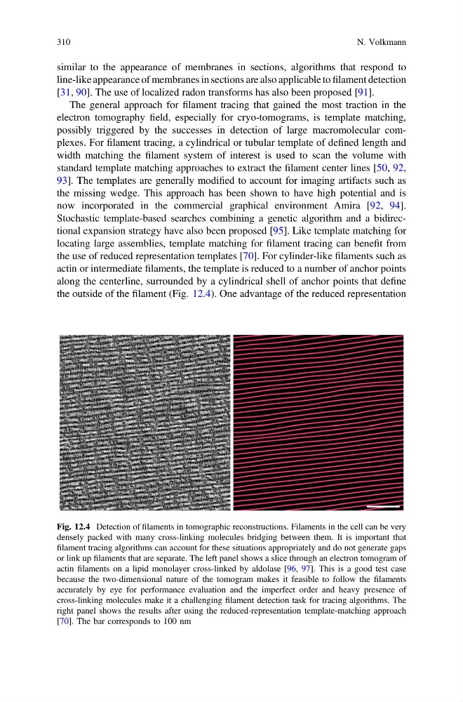
the outside of the filament (Fig. 12.4). One advantage of the reduced representation
Fig. 12.4 Detection of filaments in tomographic reconstructions. Filaments in the cell can be very
densely packed with many cross-linking molecules bridging between them. It is important that
filament tracing algorithms can account for these situations appropriately and do not generate gaps
or link up filaments that are separate. The left panel shows a slice through an electron tomogram of
actin filaments on a lipid monolayer cross-linked by aldolase [96, 97]. This is a good test case
because the two-dimensional nature of the tomogram makes it feasible to follow the filaments
accurately by eye for performance evaluation and the imperfect order and heavy presence of
cross-linking molecules make it a challenging filament detection task for tracing algorithms. The
right panel shows the results after using the reduced-representation template-matching approach
[70]. The bar corresponds to 100 nm
310
N. Volkmann
line-like appearance of membranes in sections are also applicable to filament detection
[31, 90]. The use of localized radon transforms has also been proposed [91].
The general approach for filament tracing that gained the most traction in the
electron tomography field, especially for cryo-tomograms, is template matching,
possibly triggered by the successes in detection of large macromolecular complexes. For filament tracing, a cylindrical or tubular template of defined length and
width matching the filament system of interest is used to scan the volume with
standard template matching approaches to extract the filament center lines [50, 92,
93]. The templates are generally modified to account for imaging artifacts such as
the missing wedge. This approach has been shown to have high potential and is
now incorporated in the commercial graphical environment Amira [92, 94].
Stochastic template-based searches combining a genetic algorithm and a bidirectional expansion strategy have also been proposed [95]. Like template matching for
locating large assemblies, template matching for filament tracing can benefit from
the use of reduced representation templates [70]. For cylinder-like filaments such as
actin or intermediate filaments, the template is reduced to a number of anchor points
along the centerline, surrounded by a cylindrical shell of anchor points that define
the outside of the filament (Fig. 12.4). One advantage of the reduced representation
Fig. 12.4 Detection of filaments in tomographic reconstructions. Filaments in the cell can be very
densely packed with many cross-linking molecules bridging between them. It is important that
filament tracing algorithms can account for these situations appropriately and do not generate gaps
or link up filaments that are separate. The left panel shows a slice through an electron tomogram of
actin filaments on a lipid monolayer cross-linked by aldolase [96, 97]. This is a good test case
because the two-dimensional nature of the tomogram makes it feasible to follow the filaments
accurately by eye for performance evaluation and the imperfect order and heavy presence of
cross-linking molecules make it a challenging filament detection task for tracing algorithms. The
right panel shows the results after using the reduced-representation template-matching approach
[70]. The bar corresponds to 100 nm
310
N. Volkmann