

models alone have also been used with encouraging results. Templates for different
types of membranes have allowed to distinguish between coated and uncoated
membrane patches [49, 50]. So far, these studies were limited to stained, plastic
embedded sections and may be less effective in low signal-to-noise ratio situations as
encountered in cryo-tomograms.
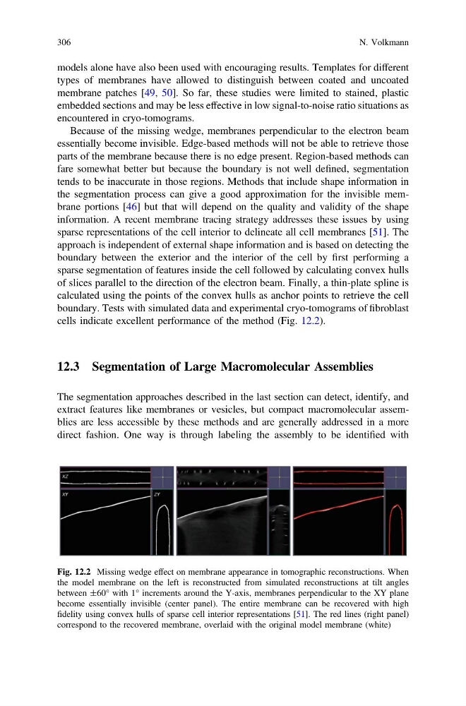
Because of the missing wedge, membranes perpendicular to the electron beam
essentially become invisible. Edge-based methods will not be able to retrieve those
parts of the membrane because there is no edge present. Region-based methods can
fare somewhat better but because the boundary is not well defined, segmentation
tends to be inaccurate in those regions. Methods that include shape information in
the segmentation process can give a good approximation for the invisible membrane portions [46] but that will depend on the quality and validity of the shape
information. A recent membrane tracing strategy addresses these issues by using
sparse representations of the cell interior to delineate all cell membranes [51]. The
approach is independent of external shape information and is based on detecting the
boundary between the exterior and the interior of the cell by first performing a
sparse segmentation of features inside the cell followed by calculating convex hulls
of slices parallel to the direction of the electron beam. Finally, a thin-plate spline is
calculated using the points of the convex hulls as anchor points to retrieve the cell
boundary. Tests with simulated data and experimental cryo-tomograms of fibroblast
cells indicate excellent performance of the method (Fig. 12.2).
12.3 Segmentation of Large Macromolecular Assemblies
The segmentation approaches described in the last section can detect, identify, and
extract features like membranes or vesicles, but compact macromolecular assemblies are less accessible by these methods and are generally addressed in a more
direct fashion. One way is through labeling the assembly to be identified with
Fig. 12.2 Missing wedge effect on membrane appearance in tomographic reconstructions. When
the model membrane on the left is reconstructed from simulated reconstructions at tilt angles
between ±60° with 1° increments around the Y-axis, membranes perpendicular to the XY plane
become essentially invisible (center panel). The entire membrane can be recovered with high
fidelity using convex hulls of sparse cell interior representations [51]. The red lines (right panel)
correspond to the recovered membrane, overlaid with the original model membrane (white)
306
N. Volkmann
types of membranes have allowed to distinguish between coated and uncoated
membrane patches [49, 50]. So far, these studies were limited to stained, plastic
embedded sections and may be less effective in low signal-to-noise ratio situations as
encountered in cryo-tomograms.
Because of the missing wedge, membranes perpendicular to the electron beam
essentially become invisible. Edge-based methods will not be able to retrieve those
parts of the membrane because there is no edge present. Region-based methods can
fare somewhat better but because the boundary is not well defined, segmentation
tends to be inaccurate in those regions. Methods that include shape information in
the segmentation process can give a good approximation for the invisible membrane portions [46] but that will depend on the quality and validity of the shape
information. A recent membrane tracing strategy addresses these issues by using
sparse representations of the cell interior to delineate all cell membranes [51]. The
approach is independent of external shape information and is based on detecting the
boundary between the exterior and the interior of the cell by first performing a
sparse segmentation of features inside the cell followed by calculating convex hulls
of slices parallel to the direction of the electron beam. Finally, a thin-plate spline is
calculated using the points of the convex hulls as anchor points to retrieve the cell
boundary. Tests with simulated data and experimental cryo-tomograms of fibroblast
cells indicate excellent performance of the method (Fig. 12.2).
12.3 Segmentation of Large Macromolecular Assemblies
The segmentation approaches described in the last section can detect, identify, and
extract features like membranes or vesicles, but compact macromolecular assemblies are less accessible by these methods and are generally addressed in a more
direct fashion. One way is through labeling the assembly to be identified with
Fig. 12.2 Missing wedge effect on membrane appearance in tomographic reconstructions. When
the model membrane on the left is reconstructed from simulated reconstructions at tilt angles
between ±60° with 1° increments around the Y-axis, membranes perpendicular to the XY plane
become essentially invisible (center panel). The entire membrane can be recovered with high
fidelity using convex hulls of sparse cell interior representations [51]. The red lines (right panel)
correspond to the recovered membrane, overlaid with the original model membrane (white)
306
N. Volkmann