

used to generate convincing segmentation results for several types of membranes
[48]. However, the current implementation requires a rough manual segmentation in
the form of manually traced slices to define the shape model and has only been
applied to situations with relatively high signal-to-noise ratios. Approaches based on
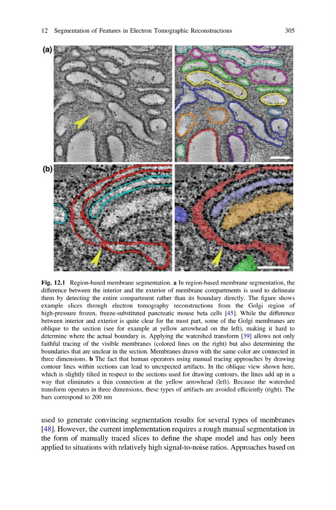
Fig. 12.1 Region-based membrane segmentation. a In region-based membrane segmentation, the
difference between the interior and the exterior of membrane compartments is used to delineate
them by detecting the entire compartment rather than its boundary directly. The figure shows
example slices through electron tomography reconstructions from the Golgi region of
high-pressure frozen, freeze-substituted pancreatic mouse beta cells [45]. While the difference
between interior and exterior is quite clear for the most part, some of the Golgi membranes are
oblique to the section (see for example at yellow arrowhead on the left), making it hard to
determine where the actual boundary is. Applying the watershed transform [39] allows not only
faithful tracing of the visible membranes (colored lines on the right) but also determining the
boundaries that are unclear in the section. Membranes drawn with the same color are connected in
three dimensions. b The fact that human operators using manual tracing approaches by drawing
contour lines within sections can lead to unexpected artifacts. In the oblique view shown here,
which is slightly tilted in respect to the sections used for drawing contours, the lines add up in a
way that eliminates a thin connection at the yellow arrowhead (left). Because the watershed
transform operates in three dimensions, these types of artifacts are avoided efficiently (right). The
bars correspond to 200 nm
12 Segmentation of Features in Electron Tomographic Reconstructions
305
[48]. However, the current implementation requires a rough manual segmentation in
the form of manually traced slices to define the shape model and has only been
applied to situations with relatively high signal-to-noise ratios. Approaches based on
Fig. 12.1 Region-based membrane segmentation. a In region-based membrane segmentation, the
difference between the interior and the exterior of membrane compartments is used to delineate
them by detecting the entire compartment rather than its boundary directly. The figure shows
example slices through electron tomography reconstructions from the Golgi region of
high-pressure frozen, freeze-substituted pancreatic mouse beta cells [45]. While the difference
between interior and exterior is quite clear for the most part, some of the Golgi membranes are
oblique to the section (see for example at yellow arrowhead on the left), making it hard to
determine where the actual boundary is. Applying the watershed transform [39] allows not only
faithful tracing of the visible membranes (colored lines on the right) but also determining the
boundaries that are unclear in the section. Membranes drawn with the same color are connected in
three dimensions. b The fact that human operators using manual tracing approaches by drawing
contour lines within sections can lead to unexpected artifacts. In the oblique view shown here,
which is slightly tilted in respect to the sections used for drawing contours, the lines add up in a
way that eliminates a thin connection at the yellow arrowhead (left). Because the watershed
transform operates in three dimensions, these types of artifacts are avoided efficiently (right). The
bars correspond to 200 nm
12 Segmentation of Features in Electron Tomographic Reconstructions
305