

11.5 Conclusion
In principle, signal optimization is a goal in all aspects of the experimental design as
well as in the reconstruction methods used to calculate volumes from the tilt-series
of projection images. However, interpretation of the tomogram often benefits from
signal optimization or filtering post-reconstruction. The need arises because of the
low contrast and noise of the reconstruction in the absence of possibilities for image
or volume averaging as in single particle reconstruction or sub-tomogram averaging. The choice of procedure may depend on the specific structural problem or the
scale of its features. In many cases a Fourier or real space filter may be sufficient to
remove high-resolution noise, such as those that are subject to artefactual interpretation. However, the signal-optimization procedure should not introduce artefacts. An objective procedure for discriminating real features from artefacts is an
important goal for which at present there is no general recipe. In real space, blurring
with a Gaussian is considered the safest method as it will not create spurious
“extrema’’ in the image or tomogram, but of course results in the loss of
high-resolution features. Optimal combination of pre-and post processing filters for
each reconstruction method is a continuing area of research.
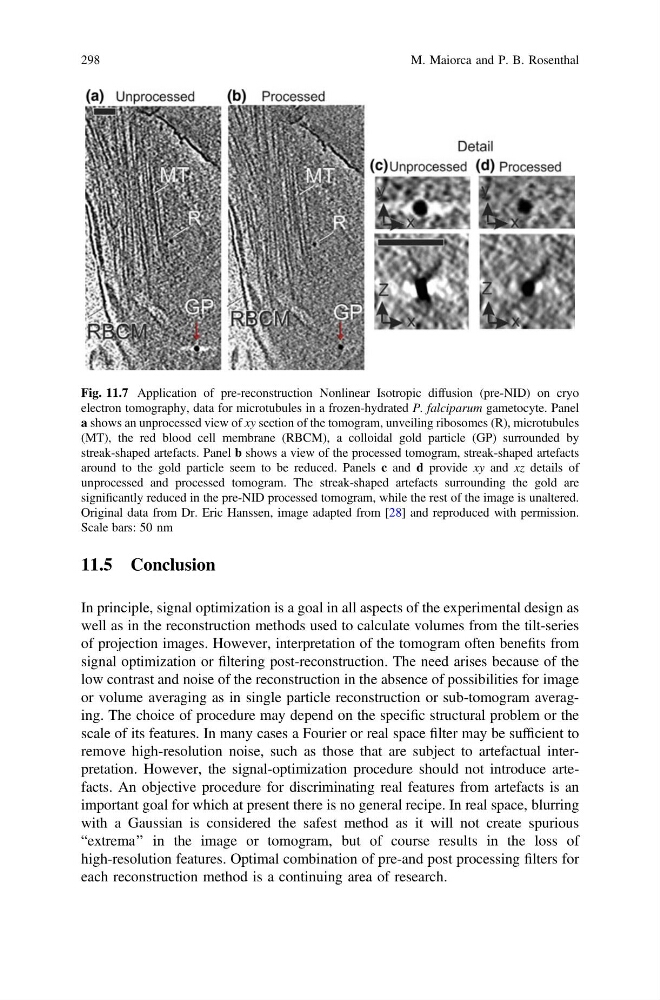
Fig. 11.7 Application of pre-reconstruction Nonlinear Isotropic diffusion (pre-NID) on cryo
electron tomography, data for microtubules in a frozen-hydrated P. falciparum gametocyte. Panel
a shows an unprocessed view of xy section of the tomogram, unveiling ribosomes (R), microtubules
(MT), the red blood cell membrane (RBCM), a colloidal gold particle (GP) surrounded by
streak-shaped artefacts. Panel b shows a view of the processed tomogram, streak-shaped artefacts
around to the gold particle seem to be reduced. Panels c and d provide xy and xz details of
unprocessed and processed tomogram. The streak-shaped artefacts surrounding the gold are
significantly reduced in the pre-NID processed tomogram, while the rest of the image is unaltered.
Original data from Dr. Eric Hanssen, image adapted from [28] and reproduced with permission.
Scale bars: 50 nm
298
M. Maiorca and P. B. Rosenthal
In principle, signal optimization is a goal in all aspects of the experimental design as
well as in the reconstruction methods used to calculate volumes from the tilt-series
of projection images. However, interpretation of the tomogram often benefits from
signal optimization or filtering post-reconstruction. The need arises because of the
low contrast and noise of the reconstruction in the absence of possibilities for image
or volume averaging as in single particle reconstruction or sub-tomogram averaging. The choice of procedure may depend on the specific structural problem or the
scale of its features. In many cases a Fourier or real space filter may be sufficient to
remove high-resolution noise, such as those that are subject to artefactual interpretation. However, the signal-optimization procedure should not introduce artefacts. An objective procedure for discriminating real features from artefacts is an
important goal for which at present there is no general recipe. In real space, blurring
with a Gaussian is considered the safest method as it will not create spurious
“extrema’’ in the image or tomogram, but of course results in the loss of
high-resolution features. Optimal combination of pre-and post processing filters for
each reconstruction method is a continuing area of research.
Fig. 11.7 Application of pre-reconstruction Nonlinear Isotropic diffusion (pre-NID) on cryo
electron tomography, data for microtubules in a frozen-hydrated P. falciparum gametocyte. Panel
a shows an unprocessed view of xy section of the tomogram, unveiling ribosomes (R), microtubules
(MT), the red blood cell membrane (RBCM), a colloidal gold particle (GP) surrounded by
streak-shaped artefacts. Panel b shows a view of the processed tomogram, streak-shaped artefacts
around to the gold particle seem to be reduced. Panels c and d provide xy and xz details of
unprocessed and processed tomogram. The streak-shaped artefacts surrounding the gold are
significantly reduced in the pre-NID processed tomogram, while the rest of the image is unaltered.
Original data from Dr. Eric Hanssen, image adapted from [28] and reproduced with permission.
Scale bars: 50 nm
298
M. Maiorca and P. B. Rosenthal