

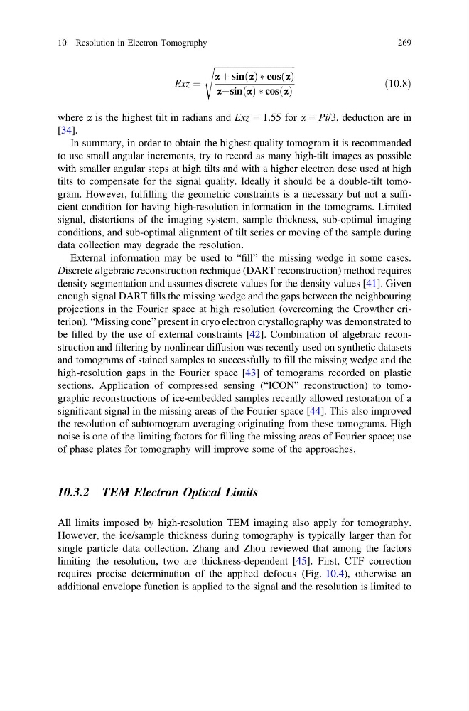
Exz ¼
ffiffiffiffiffiffiffiffiffiffiffiffiffiffiffiffiffiffiffiffiffiffiffiffiffiffiffiffiffiffiffiffiffiffiffiffi ffi
a þ sin a
ð Þ Ã cos a
ð Þ
aÀsin a
ð Þ Ã cos a
ð Þ
s
ð10:8Þ
where a is the highest tilt in radians and Exz = 1.55 for a = Pi/3, deduction are in
[34].
In summary, in order to obtain the highest-quality tomogram it is recommended
to use small angular increments, try to record as many high-tilt images as possible
with smaller angular steps at high tilts and with a higher electron dose used at high
tilts to compensate for the signal quality. Ideally it should be a double-tilt tomogram. However, fulfilling the geometric constraints is a necessary but not a sufficient condition for having high-resolution information in the tomograms. Limited
signal, distortions of the imaging system, sample thickness, sub-optimal imaging
conditions, and sub-optimal alignment of tilt series or moving of the sample during
data collection may degrade the resolution.
External information may be used to “fill” the missing wedge in some cases.
Discrete algebraic reconstruction technique (DART reconstruction) method requires
density segmentation and assumes discrete values for the density values [41]. Given
enough signal DART fills the missing wedge and the gaps between the neighbouring
projections in the Fourier space at high resolution (overcoming the Crowther criterion). “Missing cone” present in cryo electron crystallography was demonstrated to
be filled by the use of external constraints [42]. Combination of algebraic reconstruction and filtering by nonlinear diffusion was recently used on synthetic datasets
and tomograms of stained samples to successfully to fill the missing wedge and the
high-resolution gaps in the Fourier space [43] of tomograms recorded on plastic
sections. Application of compressed sensing (“ICON” reconstruction) to tomographic reconstructions of ice-embedded samples recently allowed restoration of a
significant signal in the missing areas of the Fourier space [44]. This also improved
the resolution of subtomogram averaging originating from these tomograms. High
noise is one of the limiting factors for filling the missing areas of Fourier space; use
of phase plates for tomography will improve some of the approaches.
10.3.2 TEM Electron Optical Limits
All limits imposed by high-resolution TEM imaging also apply for tomography.
However, the ice/sample thickness during tomography is typically larger than for
single particle data collection. Zhang and Zhou reviewed that among the factors
limiting the resolution, two are thickness-dependent [45]. First, CTF correction
requires precise determination of the applied defocus (Fig. 10.4), otherwise an
additional envelope function is applied to the signal and the resolution is limited to
10 Resolution in Electron Tomography
269
ffiffiffiffiffiffiffiffiffiffiffiffiffiffiffiffiffiffiffiffiffiffiffiffiffiffiffiffiffiffiffiffiffiffiffiffi ffi
a þ sin a
ð Þ Ã cos a
ð Þ
aÀsin a
ð Þ Ã cos a
ð Þ
s
ð10:8Þ
where a is the highest tilt in radians and Exz = 1.55 for a = Pi/3, deduction are in
[34].
In summary, in order to obtain the highest-quality tomogram it is recommended
to use small angular increments, try to record as many high-tilt images as possible
with smaller angular steps at high tilts and with a higher electron dose used at high
tilts to compensate for the signal quality. Ideally it should be a double-tilt tomogram. However, fulfilling the geometric constraints is a necessary but not a sufficient condition for having high-resolution information in the tomograms. Limited
signal, distortions of the imaging system, sample thickness, sub-optimal imaging
conditions, and sub-optimal alignment of tilt series or moving of the sample during
data collection may degrade the resolution.
External information may be used to “fill” the missing wedge in some cases.
Discrete algebraic reconstruction technique (DART reconstruction) method requires
density segmentation and assumes discrete values for the density values [41]. Given
enough signal DART fills the missing wedge and the gaps between the neighbouring
projections in the Fourier space at high resolution (overcoming the Crowther criterion). “Missing cone” present in cryo electron crystallography was demonstrated to
be filled by the use of external constraints [42]. Combination of algebraic reconstruction and filtering by nonlinear diffusion was recently used on synthetic datasets
and tomograms of stained samples to successfully to fill the missing wedge and the
high-resolution gaps in the Fourier space [43] of tomograms recorded on plastic
sections. Application of compressed sensing (“ICON” reconstruction) to tomographic reconstructions of ice-embedded samples recently allowed restoration of a
significant signal in the missing areas of the Fourier space [44]. This also improved
the resolution of subtomogram averaging originating from these tomograms. High
noise is one of the limiting factors for filling the missing areas of Fourier space; use
of phase plates for tomography will improve some of the approaches.
10.3.2 TEM Electron Optical Limits
All limits imposed by high-resolution TEM imaging also apply for tomography.
However, the ice/sample thickness during tomography is typically larger than for
single particle data collection. Zhang and Zhou reviewed that among the factors
limiting the resolution, two are thickness-dependent [45]. First, CTF correction
requires precise determination of the applied defocus (Fig. 10.4), otherwise an
additional envelope function is applied to the signal and the resolution is limited to
10 Resolution in Electron Tomography
269