

The effect is an apparent elongation of dense features along the electron axis and the
apparent lack of horizontal features like cellular membranes.
In order to minimize the size of the missing wedge higher tilts may be collected.
Dual-axis tomography minimizes the missing wedge to a smaller missing pyramid
[35] by recording an additional tomogram with a tilting axis X perpendicular to the
Y axis. Conical tomography [36, 37] involves tilting the sample to a high angle
(40°–60°) along one axis and rotating by a small angular step around the own axis.
Conical tomography results in an isotropic resolution in X-Y plane and a missing
cone of information in Fourier space, however is rarely used due to technical
reasons. Finally, specially designed holders in combination with FIB-milling of thin
“needles” in principle allow “on axis tilt tomography” without the missing wedge
[38]. This however may be achieved only for the samples that would form needles
highly stable during tilt series acquisition.
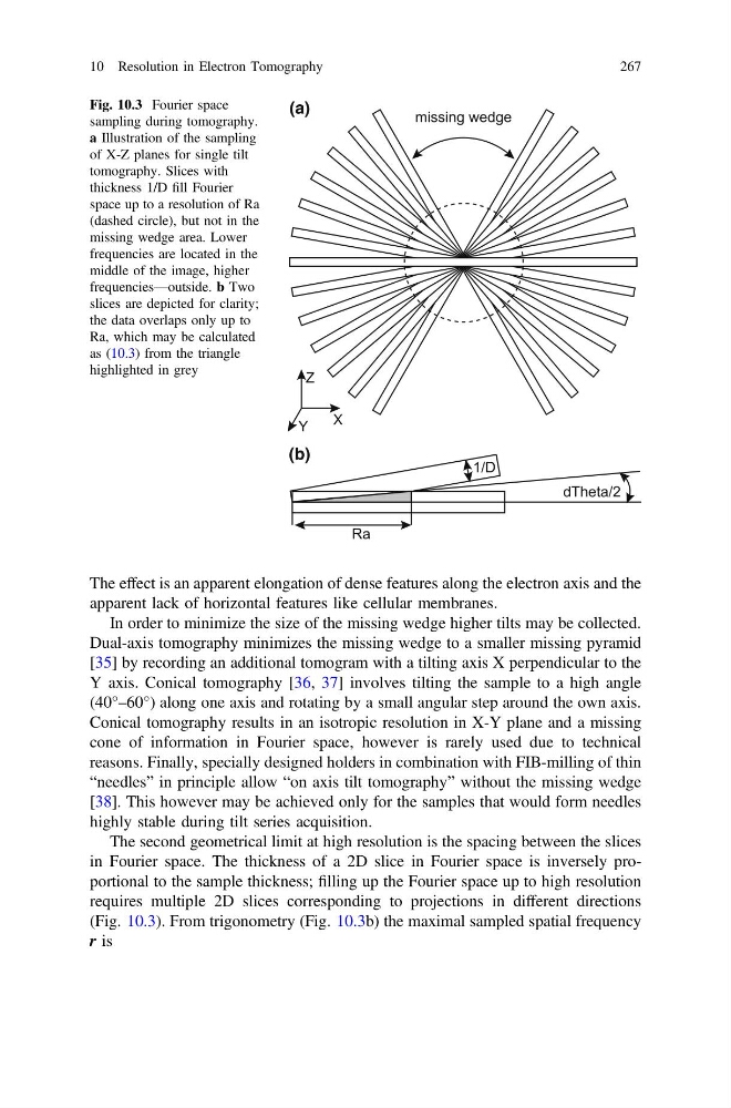
The second geometrical limit at high resolution is the spacing between the slices
in Fourier space. The thickness of a 2D slice in Fourier space is inversely proportional to the sample thickness; filling up the Fourier space up to high resolution
requires multiple 2D slices corresponding to projections in different directions
(Fig. 10.3). From trigonometry (Fig. 10.3b) the maximal sampled spatial frequency
r is
(a)
(b)
Fig. 10.3 Fourier space
sampling during tomography.
a Illustration of the sampling
of X-Z planes for single tilt
tomography. Slices with
thickness 1/D fill Fourier
space up to a resolution of Ra
(dashed circle), but not in the
missing wedge area. Lower
frequencies are located in the
middle of the image, higher
frequencies—outside. b Two
slices are depicted for clarity;
the data overlaps only up to
Ra, which may be calculated
as (10.3) from the triangle
highlighted in grey
10 Resolution in Electron Tomography
267
apparent lack of horizontal features like cellular membranes.
In order to minimize the size of the missing wedge higher tilts may be collected.
Dual-axis tomography minimizes the missing wedge to a smaller missing pyramid
[35] by recording an additional tomogram with a tilting axis X perpendicular to the
Y axis. Conical tomography [36, 37] involves tilting the sample to a high angle
(40°–60°) along one axis and rotating by a small angular step around the own axis.
Conical tomography results in an isotropic resolution in X-Y plane and a missing
cone of information in Fourier space, however is rarely used due to technical
reasons. Finally, specially designed holders in combination with FIB-milling of thin
“needles” in principle allow “on axis tilt tomography” without the missing wedge
[38]. This however may be achieved only for the samples that would form needles
highly stable during tilt series acquisition.
The second geometrical limit at high resolution is the spacing between the slices
in Fourier space. The thickness of a 2D slice in Fourier space is inversely proportional to the sample thickness; filling up the Fourier space up to high resolution
requires multiple 2D slices corresponding to projections in different directions
(Fig. 10.3). From trigonometry (Fig. 10.3b) the maximal sampled spatial frequency
r is
(a)
(b)
Fig. 10.3 Fourier space
sampling during tomography.
a Illustration of the sampling
of X-Z planes for single tilt
tomography. Slices with
thickness 1/D fill Fourier
space up to a resolution of Ra
(dashed circle), but not in the
missing wedge area. Lower
frequencies are located in the
middle of the image, higher
frequencies—outside. b Two
slices are depicted for clarity;
the data overlaps only up to
Ra, which may be calculated
as (10.3) from the triangle
highlighted in grey
10 Resolution in Electron Tomography
267