

If during an imaging workflow several factors limit the resolution, then the final
resolution will be worse than the worst of the limits:
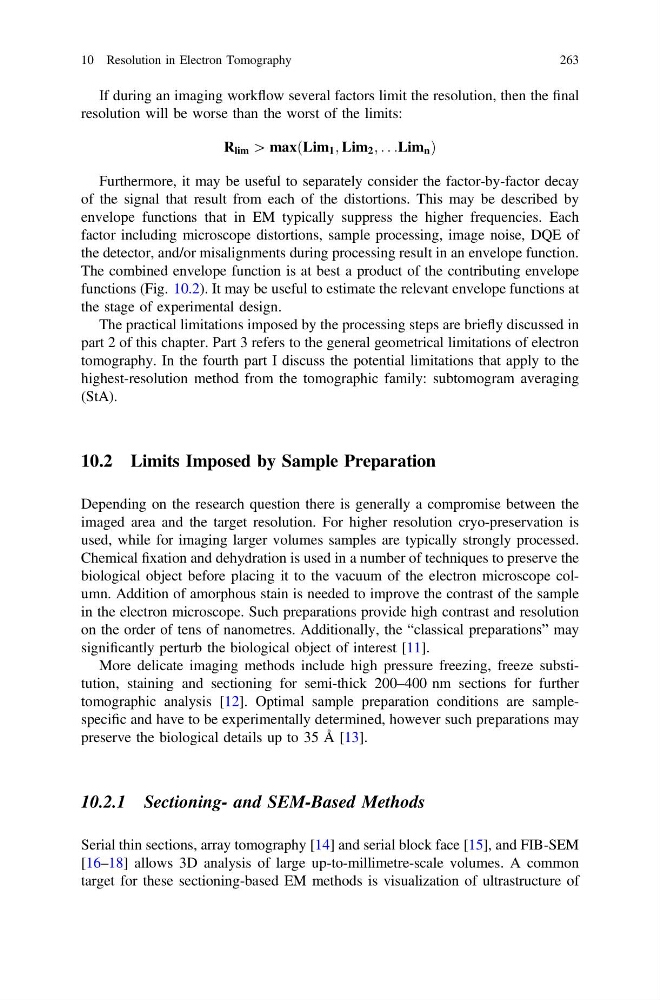
R lim [ maxðLim 1 ; Lim 2 ; . . .Lim n Þ
Furthermore, it may be useful to separately consider the factor-by-factor decay
of the signal that result from each of the distortions. This may be described by
envelope functions that in EM typically suppress the higher frequencies. Each
factor including microscope distortions, sample processing, image noise, DQE of
the detector, and/or misalignments during processing result in an envelope function.
The combined envelope function is at best a product of the contributing envelope
functions (Fig. 10.2). It may be useful to estimate the relevant envelope functions at
the stage of experimental design.
The practical limitations imposed by the processing steps are briefly discussed in
part 2 of this chapter. Part 3 refers to the general geometrical limitations of electron
tomography. In the fourth part I discuss the potential limitations that apply to the
highest-resolution method from the tomographic family: subtomogram averaging
(StA).
10.2 Limits Imposed by Sample Preparation
Depending on the research question there is generally a compromise between the
imaged area and the target resolution. For higher resolution cryo-preservation is
used, while for imaging larger volumes samples are typically strongly processed.
Chemical fixation and dehydration is used in a number of techniques to preserve the
biological object before placing it to the vacuum of the electron microscope column. Addition of amorphous stain is needed to improve the contrast of the sample
in the electron microscope. Such preparations provide high contrast and resolution
on the order of tens of nanometres. Additionally, the “classical preparations” may
significantly perturb the biological object of interest [11].
More delicate imaging methods include high pressure freezing, freeze substitution, staining and sectioning for semi-thick 200–400 nm sections for further
tomographic analysis [12]. Optimal sample preparation conditions are samplespecific and have to be experimentally determined, however such preparations may
preserve the biological details up to 35 Å [13].
10.2.1 Sectioning- and SEM-Based Methods
Serial thin sections, array tomography [14] and serial block face [15], and FIB-SEM
[16–18] allows 3D analysis of large up-to-millimetre-scale volumes. A common
target for these sectioning-based EM methods is visualization of ultrastructure of
10 Resolution in Electron Tomography
263
resolution will be worse than the worst of the limits:
R lim [ maxðLim 1 ; Lim 2 ; . . .Lim n Þ
Furthermore, it may be useful to separately consider the factor-by-factor decay
of the signal that result from each of the distortions. This may be described by
envelope functions that in EM typically suppress the higher frequencies. Each
factor including microscope distortions, sample processing, image noise, DQE of
the detector, and/or misalignments during processing result in an envelope function.
The combined envelope function is at best a product of the contributing envelope
functions (Fig. 10.2). It may be useful to estimate the relevant envelope functions at
the stage of experimental design.
The practical limitations imposed by the processing steps are briefly discussed in
part 2 of this chapter. Part 3 refers to the general geometrical limitations of electron
tomography. In the fourth part I discuss the potential limitations that apply to the
highest-resolution method from the tomographic family: subtomogram averaging
(StA).
10.2 Limits Imposed by Sample Preparation
Depending on the research question there is generally a compromise between the
imaged area and the target resolution. For higher resolution cryo-preservation is
used, while for imaging larger volumes samples are typically strongly processed.
Chemical fixation and dehydration is used in a number of techniques to preserve the
biological object before placing it to the vacuum of the electron microscope column. Addition of amorphous stain is needed to improve the contrast of the sample
in the electron microscope. Such preparations provide high contrast and resolution
on the order of tens of nanometres. Additionally, the “classical preparations” may
significantly perturb the biological object of interest [11].
More delicate imaging methods include high pressure freezing, freeze substitution, staining and sectioning for semi-thick 200–400 nm sections for further
tomographic analysis [12]. Optimal sample preparation conditions are samplespecific and have to be experimentally determined, however such preparations may
preserve the biological details up to 35 Å [13].
10.2.1 Sectioning- and SEM-Based Methods
Serial thin sections, array tomography [14] and serial block face [15], and FIB-SEM
[16–18] allows 3D analysis of large up-to-millimetre-scale volumes. A common
target for these sectioning-based EM methods is visualization of ultrastructure of
10 Resolution in Electron Tomography
263